Characterization of Breast Lesions: Comparison of Digital Breast Tomosynthesis and Ultrasonography
- Affiliations
-
- 1Department of Radiology, Human Medical Imaging & Intervention Center, Seoul 135-120, Korea.
- 2Department of Radiology, Seoul National University Hospital, Seoul 110-744, Korea. imchangjm@gmail.com
- 3Department of Radiology, Seoul National University Hospital Healthcare System Gangnam Center, Seoul 135-984, Korea.
- KMID: 2070169
- DOI: http://doi.org/10.3348/kjr.2015.16.2.229
Abstract
OBJECTIVE
To compare the diagnostic performance of digital breast tomosynthesis (DBT) and conventional breast ultrasound (US) to characterize breast lesions as benign or malignant.
MATERIALS AND METHODS
A total of 332 women, presenting for screening examinations or for breast biopsy between March and June 2012 were recruited to undergo digital mammography (DM), DBT, and breast US examination. Among them, 113 patients with 119 breast lesions depicted on DM were finally included. Three blinded radiologists performed an enriched reader study and reviewed the DBT and US images. Each reader analyzed the lesions in random order, assigned Breast Imaging Reporting and Data System (BI-RADS) descriptors, rated the images for the likelihood of malignancy (%) and made a BI-RADS final assessment. Diagnostic accuracy, as assessed by the area under the receiver operating characteristic curve, sensitivity, and specificity of DBT and US were compared.
RESULTS
Among the 119 breast lesions depicted on DM, 75 were malignant and the remaining 44 were benign. The average diagnostic performance for characterizing breast lesions as benign or malignant in terms of area under the curve was 0.899 for DBT and 0.914 for US (p = 0.394). Mean sensitivity (97.3% vs. 98.7%, p = 0.508) and specificity (44.7% vs. 39.4%, p = 0.360) were also not significantly different.
CONCLUSION
Digital breast tomosynthesis may provide similar reader lesion characterization performance to that of US for breast lesions depicted on DM.
MeSH Terms
Figure
-
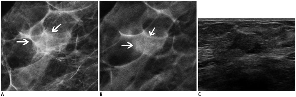
Fig. 1 62-year-old woman with 1.2 cm-sized invasive ductal carcinoma. A. Craniocaudal digital mammography view. B. Craniocaudal digital breast tomosynthesis (DBT) view (1-mm section). C. Transverse ultrasonography (US) view. Oval isodense mass (arrows) with indistinct margins is observed on DBT. This lesion appears as hypoechoic lesion with angular margins on US. Readers 1 and 3 categorized this lesion as Breast Imaging Reporting and Data System (BI-RADS) category 3, and reader 2 categorized this lesion as BI-RADS category 4A on DBT. Readers 1 and 2 categorized this lesion as BI-RADS category 4C, and reader 3 categorized this lesion as BI-RADS category 4A on US.
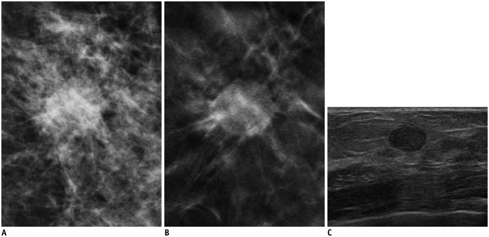
Fig. 2 57-year-old woman with 1.2 cm-sized invasive papillary carcinoma. A. Craniocaudal full-field digital mammography view. B. Craniocaudal digital breast tomosynthesis (DBT) (1-mm section) view. C. Transverse ultrasonography (US) view. Mass with spiculated margin is clearly visible on DBT image. This lesion appears as oval circumscribed lesion on US. Readers 1 and 2 categorized this lesion as Breast Imaging Reporting and Data System (BI-RADS) category 4B, and reader 3 categorized this lesion as BI-RADS category 4A on DBT. Readers 2 and 3 categorized this lesion as BI-RADS category 3, and reader 1 categorized this lesion as BI-RADS category 4A on US.
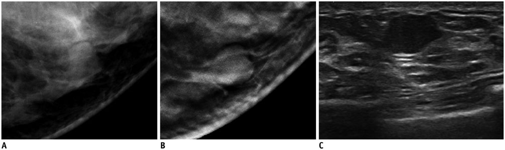
Fig. 3 63-year-old woman with fibroadenoma (1.3 cm in diameter as measured on ultrasonography [US]). A. Mediolateral oblique digital mammography view. B. Mediolateral oblique digital breast tomosynthesis (DBT) (1-mm section) view. C. Transverse US view. Round isodense mass with circumscribed margins is observed on DBT. This lesion appears as irregular hypoechoic circumscribed mass on US. Readers 1 and 2 categorized this lesion as Breast Imaging Reporting and Data System (BI-RADS) category 3, and reader 3 categorized this lesion as BI-RADS category 2 on DBT. Readers 1 and 2 categorized this lesion as BI-RADS category 4A, and reader 3 categorized this lesion as BI-RADS category 3 on US.
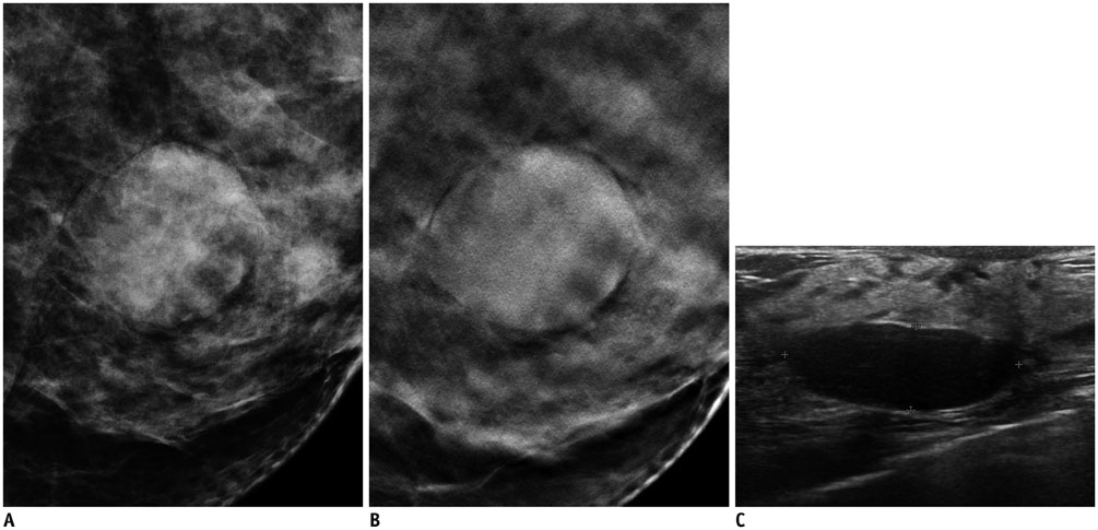
Fig. 4 38-year-old woman with 2.5-cm benign phyllodes tumor. A. Mediolateral oblique digital mammography view. B. Mediolateral oblique digital breast tomosynthesis (DBT) (1-mm section) view. C. Transverse ultrasonography (US) view. Circumscribed high density mass is observed on DBT images. Mass is oval, circumscribed and accompanied by posterior acoustic enhancement on US. All readers categorized this lesion as Breast Imaging Reporting and Data System (BI-RADS) category 4A on DBT. All readers categorized this lesion as BI-RADS category 3 on US.
Cited by 4 articles
-
Addition of Digital Breast Tomosynthesis to Full-Field Digital Mammography in the Diagnostic Setting: Additional Value and Cancer Detectability
Mirinae Seo, Jung Min Chang, Sun Ah Kim, Won Hwa Kim, Ji He Lim, Su Hyun Lee, Min Sun Bae, Hye Ryoung Koo, Nariya Cho, Woo Kyung Moon
J Breast Cancer. 2016;19(4):438-446. doi: 10.4048/jbc.2016.19.4.438.Performance of Screening Mammography: A Report of the Alliance for Breast Cancer Screening in Korea
Eun Hye Lee, Keum Won Kim, Young Joong Kim, Dong-Rock Shin, Young Mi Park, Hyo Soon Lim, Jeong Seon Park, Hye-Won Kim, You Me Kim, Hye Jung Kim, Jae Kwan Jun
Korean J Radiol. 2016;17(4):489-496. doi: 10.3348/kjr.2016.17.4.489.Analysis of Participant Factors That Affect the Diagnostic Performance of Screening Mammography: A Report of the Alliance for Breast Cancer Screening in Korea
Young Joong Kim, Eun Hye Lee, Jae Kwan Jun, Dong-Rock Shin, Young Mi Park, Hye-Won Kim, Youme Kim, Keum Won Kim, Hyo Soon Lim, Jeong Seon Park, Hye Jung Kim, Hye-Mi Jo,
Korean J Radiol. 2017;18(4):624-631. doi: 10.3348/kjr.2017.18.4.624.Effect of a Deep Learning Framework-Based Computer-Aided Diagnosis System on the Diagnostic Performance of Radiologists in Differentiating between Malignant and Benign Masses on Breast Ultrasonography
Ji Soo Choi, Boo-Kyung Han, Eun Sook Ko, Jung Min Bae, Eun Young Ko, So Hee Song, Mi-ri Kwon, Jung Hee Shin, Soo Yeon Hahn
Korean J Radiol. 2019;20(5):749-758. doi: 10.3348/kjr.2018.0530.
Reference
-
1. Berry DA, Cronin KA, Plevritis SK, Fryback DG, Clarke L, Zelen M, et al. Effect of screening and adjuvant therapy on mortality from breast cancer. N Engl J Med. 2005; 353:1784–1792.2. Hellquist BN, Duffy SW, Abdsaleh S, Björneld L, Bordás P, Tabár L, et al. Effectiveness of population-based service screening with mammography for women ages 40 to 49 years: evaluation of the Swedish Mammography Screening in Young Women (SCRY) cohort. Cancer. 2011; 117:714–722.3. Tabár L, Fagerberg CJ, Gad A, Baldetorp L, Holmberg LH, Gröntoft O, et al. Reduction in mortality from breast cancer after mass screening with mammography. Randomised trial from the Breast Cancer Screening Working Group of the Swedish National Board of Health and Welfare. Lancet. 1985; 1:829–883.4. Tabar L, Yen MF, Vitak B, Chen HH, Smith RA, Duffy SW. Mammography service screening and mortality in breast cancer patients: 20-year follow-up before and after introduction of screening. Lancet. 2003; 361:1405–1410.5. Mandelson MT, Oestreicher N, Porter PL, White D, Finder CA, Taplin SH, et al. Breast density as a predictor of mammographic detection: comparison of interval- and screen-detected cancers. J Natl Cancer Inst. 2000; 92:1081–1087.6. Kerlikowske K, Grady D, Barclay J, Sickles EA, Ernster V. Effect of age, breast density, and family history on the sensitivity of first screening mammography. JAMA. 1996; 276:33–38.7. Kopans DB. Digital breast tomosynthesis from concept to clinical care. AJR Am J Roentgenol. 2014; 202:299–308.8. Niklason LT, Christian BT, Niklason LE, Kopans DB, Castleberry DE, Opsahl-Ong BH, et al. Digital tomosynthesis in breast imaging. Radiology. 1997; 205:399–406.9. Gennaro G, Toledano A, di Maggio C, Baldan E, Bezzon E, La Grassa M, et al. Digital breast tomosynthesis versus digital mammography: a clinical performance study. Eur Radiol. 2010; 20:1545–1553.10. Good WF, Abrams GS, Catullo VJ, Chough DM, Ganott MA, Hakim CM, et al. Digital breast tomosynthesis: a pilot observer study. AJR Am J Roentgenol. 2008; 190:865–869.11. Gur D, Abrams GS, Chough DM, Ganott MA, Hakim CM, Perrin RL, et al. Digital breast tomosynthesis: observer performance study. AJR Am J Roentgenol. 2009; 193:586–591.12. Spangler ML, Zuley ML, Sumkin JH, Abrams G, Ganott MA, Hakim C, et al. Detection and classification of calcifications on digital breast tomosynthesis and 2D digital mammography: a comparison. AJR Am J Roentgenol. 2011; 196:320–324.13. Tagliafico A, Astengo D, Cavagnetto F, Rosasco R, Rescinito G, Monetti F, et al. One-to-one comparison between digital spot compression view and digital breast tomosynthesis. Eur Radiol. 2012; 22:539–544.14. Teertstra HJ, Loo CE, van den Bosch MA, van Tinteren H, Rutgers EJ, Muller SH, et al. Breast tomosynthesis in clinical practice: initial results. Eur Radiol. 2010; 20:16–24.15. Noroozian M, Hadjiiski L, Rahnama-Moghadam S, Klein KA, Jeffries DO, Pinsky RW, et al. Digital breast tomosynthesis is comparable to mammographic spot views for mass characterization. Radiology. 2012; 262:61–68.16. Hakim CM, Chough DM, Ganott MA, Sumkin JH, Zuley ML, Gur D. Digital breast tomosynthesis in the diagnostic environment: a subjective side-by-side review. AJR Am J Roentgenol. 2010; 195:W172–W176.17. Frazier TG, Murphy JT, Furlong A. The selected use of ultrasound mammography to improve diagnostic accuracy in carcinoma of the breast. J Surg Oncol. 1985; 29:231–232.18. Kaplan SS. Clinical utility of bilateral whole-breast US in the evaluation of women with dense breast tissue. Radiology. 2001; 221:641–649.19. Bassett LW. Imaging of breast masses. Radiol Clin North Am. 2000; 38:669–691. vii-viii.20. Dennis MA, Parker SH, Klaus AJ, Stavros AT, Kaske TI, Clark SB. Breast biopsy avoidance: the value of normal mammograms and normal sonograms in the setting of a palpable lump. Radiology. 2001; 219:186–191.21. Graf O, Helbich TH, Hopf G, Graf C, Sickles EA. Probably benign breast masses at US: is follow-up an acceptable alternative to biopsy? Radiology. 2007; 244:87–93.22. Houssami N, Irwig L, Simpson JM, McKessar M, Blome S, Noakes J. Sydney Breast Imaging Accuracy Study: comparative sensitivity and specificity of mammography and sonography in young women with symptoms. AJR Am J Roentgenol. 2003; 180:935–940.23. D'Orsi CJ, Bassett LW, Berg WA, Feig SA, Jackson VP, Kopans DB, et al. Breast Imaging Reporting and Data System, BI-RADS: Mammography. 4th ed. Reston: American College of Radiology;2003.24. Mendelson EB, Baum JK, Berg WA, Merritt CB, Rubin E. Breast Imaging Reporting and Data System, BI-RADS: Ultrasound. 1st ed. Reston: American College of Radiology;2003.25. Rafferty EA, Park JM, Philpotts LE, Poplack SP, Sumkin JH, Halpern EF, et al. Assessing radiologist performance using combined digital mammography and breast tomosynthesis compared with digital mammography alone: results of a multicenter, multireader trial. Radiology. 2013; 266:104–113.26. Zuley ML, Bandos AI, Ganott MA, Sumkin JH, Kelly AE, Catullo VJ, et al. Digital breast tomosynthesis versus supplemental diagnostic mammographic views for evaluation of noncalcified breast lesions. Radiology. 2013; 266:89–95.27. Vercauteren LD, Kessels AG, van der Weijden T, Koster D, Severens JL, van Engelshoven JM, et al. Clinical impact of the use of additional ultrasonography in diagnostic breast imaging. Eur Radiol. 2008; 18:2076–2084.28. Flobbe K, Bosch AM, Kessels AG, Beets GL, Nelemans PJ, von Meyenfeldt MF, et al. The additional diagnostic value of ultrasonography in the diagnosis of breast cancer. Arch Intern Med. 2003; 163:1194–1199.29. Leconte I, Feger C, Galant C, Berlière M, Berg BV, D'Hoore W, et al. Mammography and subsequent whole-breast sonography of nonpalpable breast cancers: the importance of radiologic breast density. AJR Am J Roentgenol. 2003; 180:1675–1679.30. Andersson I, Ikeda DM, Zackrisson S, Ruschin M, Svahn T, Timberg P, et al. Breast tomosynthesis and digital mammography: a comparison of breast cancer visibility and BIRADS classification in a population of cancers with subtle mammographic findings. Eur Radiol. 2008; 18:2817–2825.31. Lazarus E, Mainiero MB, Schepps B, Koelliker SL, Livingston LS. BI-RADS lexicon for US and mammography: interobserver variability and positive predictive value. Radiology. 2006; 239:385–391.32. Lee SH, Chang JM, Cho N, Koo HR, Yi A, Kim SJ, et al. Practice guideline for the performance of breast ultrasound elastography. Ultrasonography. 2014; 33:3–10.
