Korean J Radiol.
2015 Feb;16(1):201-205. 10.3348/kjr.2015.16.1.201.
Assessment of Bone Age in Prepubertal Healthy Korean Children: Comparison among the Korean Standard Bone Age Chart, Greulich-Pyle Method, and Tanner-Whitehouse Method
- Affiliations
-
- 1Department of Radiology, Dankook University Hospital, Cheonan 330-715, Korea. yslee@dkuh.co.kr
- 2Department of Pediatrics, Dankook University Hospital, Cheonan 330-715, Korea.
- KMID: 2070001
- DOI: http://doi.org/10.3348/kjr.2015.16.1.201
Abstract
OBJECTIVE
To compare the reliability of the Greulich-Pyle (GP) method, Tanner-Whitehouse 3 (TW3) method and Korean standard bone age chart (KS) in the evaluation of bone age of prepubertal healthy Korean children.
MATERIALS AND METHODS
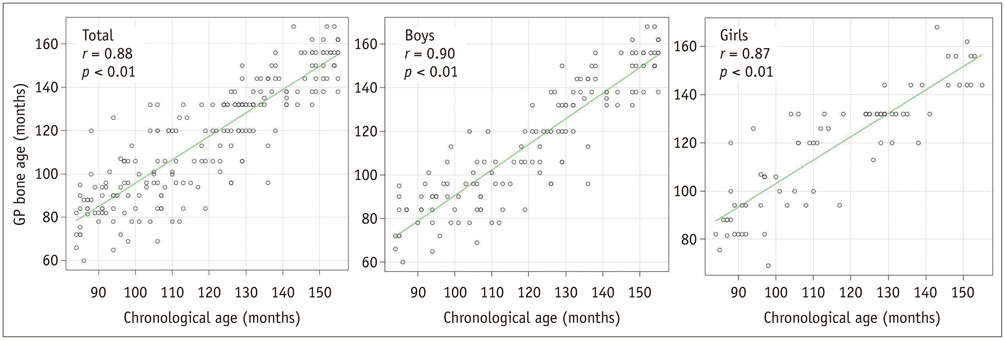
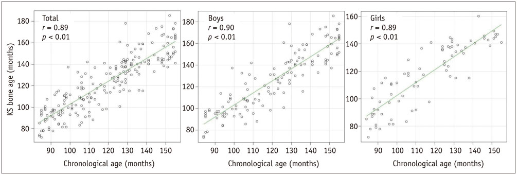
Left hand-wrist radiographs of 212 prepubertal healthy Korean children aged 7 to 12 years, obtained for the evaluation of the traumatic injury in emergency department, were analyzed by two observers. Bone age was estimated using the GP method, TW3 method and KS, and was calculated in months. The correlation between bone age measured by each method and chronological age of each child was analyzed using Pearson correlation coefficient, scatterplot. The three methods were compared using one-way analysis of variance.
RESULTS
Significant correlations were found between chronological age and bone age estimated by all three methods in whole group and in each gender (R2 ranged from 0.87 to 0.9, p < 0.01). Although bone age estimated by KS was slightly closer to chronological age than those estimated by the GP and TW3 methods, the difference between three methods was not statistically significant (p > 0.01).
CONCLUSION
The KS, GP, and TW3 methods show good reliability in the evaluation of bone age of prepubertal healthy Korean children without significant difference between them. Any are useful for evaluation of bone age in prepubertal healthy Korean children.
MeSH Terms
Figure
Cited by 3 articles
-
Prediction of endotracheal tube size for pediatric patients from the epiphysis diameter of radius
Hee Young Kim, Ji Hyun Cheon, Seung Hoon Baek, Kyung Hoon Kim, Tae Kyun Kim
Korean J Anesthesiol. 2017;70(1):52-57. doi: 10.4097/kjae.2017.70.1.52.Morning basal luteinizing hormone, a good screening tool for diagnosing central precocious puberty
Dong-Min Lee, In-Hyuk Chung
Ann Pediatr Endocrinol Metab. 2019;24(1):27-33. doi: 10.6065/apem.2019.24.1.27.Clinical validation of a deep-learning-based bone age software in healthy Korean children
Hyo-Kyoung Nam, Winnah Wu-In Lea, Zepa Yang, Eunjin Noh, Young-Jun Rhie, Kee-Hyoung Lee, Suk-Joo Hong
Ann Pediatr Endocrinol Metab. 2024;29(2):102-108. doi: 10.6065/apem.2346050.025.
Reference
-
1. Zerin JM, Hernandez RJ. Approach to skeletal maturation. Hand Clin. 1991; 7:53–62.2. Greulich WW, Pyle SI. Radiographic atlas of skeletal development of hand wrist. Stanford: Stanford University Press;1971.3. Tanner JM, Healy MJ, Goldstein H, Cameron N. Assessment of skeletal maturity and prediction of adult height (TW3 method). London: W.B Saunders Company;2001.4. Ontell FK, Ivanovic M, Ablin DS, Barlow TW. Bone age in children of diverse ethnicity. AJR Am J Roentgenol. 1996; 167:1395–1398.5. Zhang A, Sayre JW, Vachon L, Liu BJ, Huang HK. Racial differences in growth patterns of children assessed on the basis of bone age. Radiology. 2009; 250:228–235.6. Yeon KM, Kim IO. Standard atlas of bone-age in Korean children. Seoul: Sungmoonbook;1999.7. Yeon KM. Standard bone-age of infants and children in Korea. J Korean Med Sci. 1997; 12:9–16.8. Kim SY, Oh YJ, Shin JY, Rhie YJ, Lee KH. Comparison of the Greulich-Pyle and Tanner Whitehouse (TW3) methods in bone age assessment. J Korean Soc Pediatr Endocrinol. 2008; 13:50–55.9. Vignolo M, Milani S, DiBattista E, Naselli A, Mostert M, Aicardi G. Modified Greulich-Pyle, Tanner-Whitehouse, and Roche-Wainer-Thissen (knee) methods for skeletal age assessment in a group of Italian children and adolescents. Eur J Pediatr. 1990; 149:314–317.10. Cole AJ, Webb L, Cole TJ. Bone age estimation: a comparison of methods. Br J Radiol. 1988; 61:683–686.11. Milner GR, Levick RK, Kay R. Assessment of bone age: a comparison of the Greulich and Pyle, and the Tanner and Whitehouse methods. Clin Radiol. 1986; 37:119–121.12. Bull RK, Edwards PD, Kemp PM, Fry S, Hughes IA. Bone age assessment: a large scale comparison of the Greulich and Pyle, and Tanner and Whitehouse (TW2) methods. Arch Dis Child. 1999; 81:172–173.13. Ashizawa K, Asami T, Anzo M, Matsuo N, Matsuoka H, Murata M, et al. Standard RUS skeletal maturation of Tokyo children. Ann Hum Biol. 1996; 23:457–469.14. Zhen OY, Baolin L. Skeletal maturity of the hand and wrist in Chinese school children in Harbin assessed by the TW2 method. Ann Hum Biol. 1986; 13:183–187.
- Full Text Links
- Actions
-
Cited
- CITED
-
- Close
- Share
- Similar articles
-
- Assessment of Bone Age: A comparison of the Greulich Pyle Method to the Tanner Whitehouse Method
- Comparison of the Greulich-Pyle and Tanner Whitehouse (TW3) Methods in Bone Age Assessment
- Clinical Validation of a Deep Learning-Based Hybrid (Greulich-Pyle and Modified Tanner-Whitehouse) Method for Bone Age Assessment
- Comparison of Bone Ages in Early Puberty: Computerized Greulich-Pyle Based Bone Age vs. Sauvegrain Method
- Comparison of predicted adult heights measured by Bayley-Pinneau and Tanner-Whitehouse 3 methods in normal children, those with precocious puberty and with constitutional growth delay


