Charcoal-Induced Granuloma That Mimicked a Nodal Metastasis on Ultrasonography and FDG-PET/CT after Neck Dissection
- Affiliations
-
- 1Department of Radiology, Konkuk University Medical Center, Konkuk University School of Medicine, Seoul 143-729, Korea. mdmoonwj@naver.com
- 2Department of Nuclear Medicine, Konkuk University Medical Center, Konkuk University School of Medicine, Seoul 143-729, Korea.
- 3Department of Pathology, Konkuk University Medical Center, Konkuk University School of Medicine, Seoul 143-729, Korea.
- 4Department of Surgery, Konkuk University Medical Center, Konkuk University School of Medicine, Seoul 143-729, Korea.
- KMID: 2070000
- DOI: http://doi.org/10.3348/kjr.2015.16.1.196
Abstract
- Charcoal can be used for preoperative localization of metastatic lymph nodes in the neck. Charcoal remains stable without causing foreign body reactions during as hort period. However, foreign body reactions may develop if charcoal is left in situ for more than 6 months. We reported a case of charcoal granuloma mimicking local recurrence on fluorodeoxyglucose-positron emission tomography/computed tomography and ultrasonography in a 47-year-old woman who had cervical lymph node dissection due to metastatic invasive ductal carcinoma of the breast.
Keyword
MeSH Terms
-
Breast Neoplasms/pathology/surgery/therapy
Carcinoma/*pathology/surgery/therapy
Cervix Uteri/pathology/ultrasonography
Charcoal/toxicity
Female
Fluorodeoxyglucose F18/diagnostic use
Granuloma/*diagnosis/pathology
Humans
Lymph Nodes/*surgery/ultrasonography
Lymphatic Metastasis
Middle Aged
Neoplasm Recurrence, Local
Positron-Emission Tomography
Radiopharmaceuticals/diagnostic use
Tomography, X-Ray Computed
Charcoal
Fluorodeoxyglucose F18
Radiopharmaceuticals
Figure
-
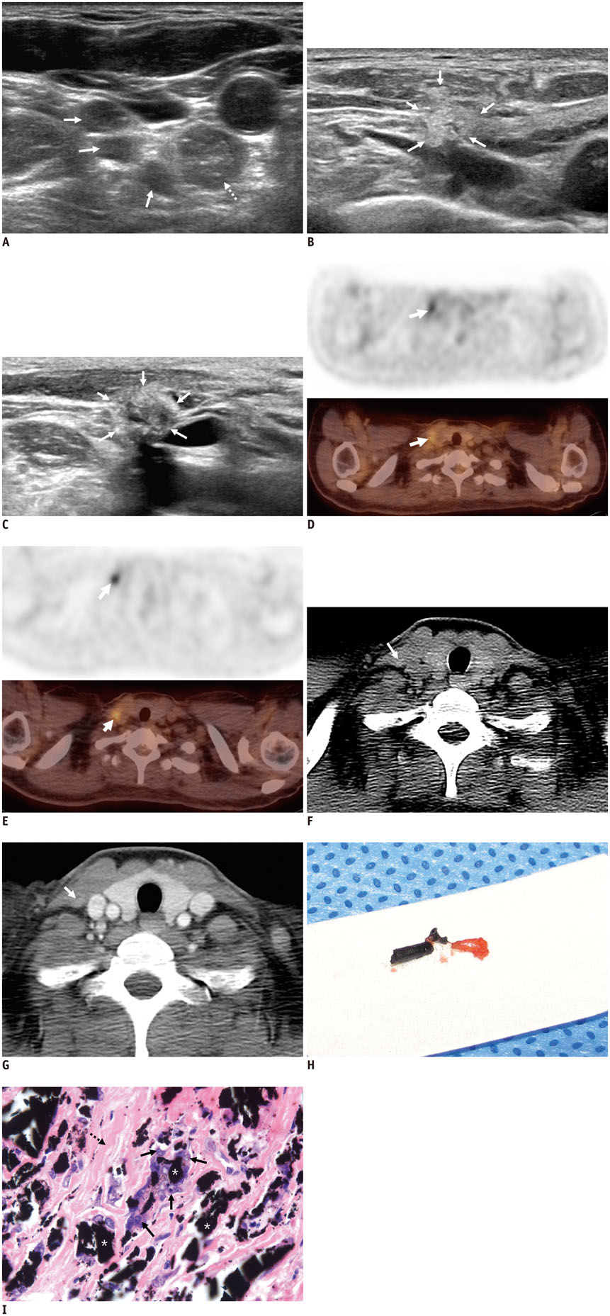
Fig. 1 US, PET/CT, chest CT and pathologic findings of charcoal-induced granuloma. Preoperative US (A) shows several hypoechoic nodules with well-defined margins (arrows) and one well-defined, isoechoic nodule (broken arrow) with internal high echoic dots in right level IV. These lymph nodes were localized using charcoal and revealed metastatic lymph nodes with perinodal infiltration. Follow-up US at 6 months (B) shows irregularly shaped, hyperechoic nodule measuring 9.8 mm with indistinct margin with adjacent sternocleidomastoid muscle and fat of posterior cervical space (arrows). On 2-year follow-up US for US-guided core biopsy (C), compared to previous US (B), echogenicity of nodule decreased, and posterior acoustic shadowing was prominent (arrows). Six-month, initial follow-up FDG-PET and PET/CT (D) shows hypermetabolic nodule (SUVmax = 4.0) at right level IV (arrow). FDG = fluorodeoxyglucose, PET = positron emission tomography, SUVmax = maximum standardized uptake value, US = ultrasonography. Lesion is still hypermetablic (SUVmax = 4.3) (arrow) on 18-month follow-up FDG-PET and PET/CT (E). Precontrast chest CT (F) acquired at same day shows slightly hyperdense, oval-shaped nodule at lateral aspect of right internal jugular vein (arrow). Postcontrast CT (G) shows no definite enhancement of nodule (arrow). Gross specimen (H) shows black-pigmented soft tissue fragment. Microscopic image (H&E staining, × 300) (I) shows black pigments suggesting charcoal particles (asterisks) surrounded by multinucleared giant cells (arrows) and fibrosis (broken arrow). FDG = fluorodeoxyglucose, PET = positron emission tomography, SUVmax = maximum standardized uptake value, US = ultrasonography
Reference
-
1. Langlois SL, Carter ML. Carbon localisation of impalpable mammographic abnormalities. Australas Radiol. 1991; 35:237–241.2. Kang TW, Shin JH, Han BK, Ko EY, Kang SS, Hahn SY, et al. Preoperative ultrasound-guided tattooing localization of recurrences after thyroidectomy: safety and effectiveness. Ann Surg Oncol. 2009; 16:1655–1659.3. Chung YE, Kim EK, Kim MJ, Yun M, Hong SW. Suture granuloma mimicking recurrent thyroid carcinoma on ultrasonography. Yonsei Med J. 2006; 47:748–751.4. Kim JH, Lee JH, Shong YK, Hong SJ, Ko MS, Lee DH, et al. Ultrasound features of suture granulomas in the thyroid bed after thyroidectomy for papillary thyroid carcinoma with an emphasis on their differentiation from locally recurrent thyroid carcinomas. Ultrasound Med Biol. 2009; 35:1452–1457.5. Saito N, Nadgir RN, Nakahira M, Takahashi M, Uchino A, Kimura F, et al. Posttreatment CT and MR imaging in head and neck cancer: what the radiologist needs to know. Radiographics. 2012; 32:1261–1282. discussion 1282-1284.6. Bonhomme-Faivre L, Depraetere P, Savelli MP, Amdidouche D, Bizi E, Seiller M, et al. Charcoal suspension for tumor labelling modifies macrophage activity in mice. Life Sci. 2000; 66:817–827.7. Ruiz-Delgado ML, López-Ruiz JA, Sáiz-López A. Abnormal mammography and sonography associated with foreign-body giant-cell reaction after stereotactic vacuum-assisted breast biopsy with carbon marking. Acta Radiol. 2008; 49:1112–1118.8. Shin JH, Han BK, Ko EY, Kang SS. Sonographic findings in the surgical bed after thyroidectomy: comparison of recurrent tumors and nonrecurrent lesions. J Ultrasound Med. 2007; 26:1359–1366.9. Kim YK, Park HS. Foreign body granuloma of activated charcoal. Abdom Imaging. 2008; 33:94–97.10. Lim ST, Jeong HJ, Kim DW, Yim CY, Sohn MH. F-18 FDG PET-CT findings of intraperitoneal carbon particles-induced granulomas mimicking peritoneal carcinomatosis. Clin Nucl Med. 2008; 33:321–324.
- Full Text Links
-
- Actions
-
Cited
- CITED
-
- Close
- Share
-
- Similar articles
-
- False Positive of F-18 FDG-PET/CT due to Activated Charcoal Granuloma from Intraperitoneal Chemotherapy: A Case Report
- The Diagnostic Utility of Ultrasonography, CT and PET/CT for the Preoperative Evaluation of Cervical Lymph Node Metastasis in Papillary Thyroid Cancer Patients
- Comparison of Neck CT and ¹â¸F-FDG PET-CT for Making the Preoperative Diagnosis of Lymph Node Metastasis in Papillary Thyroid Cancer
- A Case of Recurrence-Mimicking Charcoal Granuloma in a Breast Cancer Patient: Ultrasound, CT, PET/CT and Breast-Specific Gamma Imaging Findings
- Comparison of 18F FDG-PET and CT/MRI for the Diagnosis of Cervical Lymph Node Metastasis in Head and Neck Cancer: A Level-by-Level Based Study
