Intra-Articular Fibroma of Tendon Sheath in a Knee Joint Associated with Iliotibial Band Friction Syndrome
- Affiliations
-
- 1Department of Radiology, Dong-A University Medical Center, Busan 602-715, Korea. hdhdoc@naver.com
- 2Department of Pathology, Dong-A University Medical Center, Busan 602-715, Korea.
- 3Department of Orthopedics, Dong-A University Medical Center, Busan 602-715, Korea.
- KMID: 2069996
- DOI: http://doi.org/10.3348/kjr.2015.16.1.169
Abstract
- Iliotibial band (ITB) friction syndrome is a common overuse injury typically seen in the active athlete population. A nodular lesion on the inner side of the ITB as an etiology or an accompanying lesion from friction syndrome has been rarely reported. A 45-year-old male presented with recurrent pain and a movable nodule at the lateral joint area, diagnosed as ITB friction syndrome. The nodule was confirmed as a rare intra-articular fibroma of the tendon sheath (FTS) on the basis of histopathologic findings. We describe the MRI findings, arthroscopic and pathologic features, in this case of intra-articular FTS presenting with ITB friction syndrome.
Keyword
MeSH Terms
Figure
-
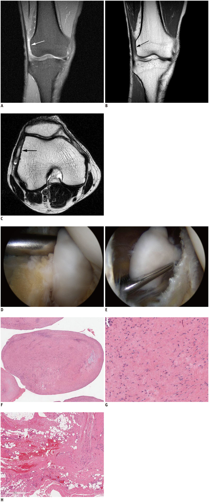
Fig. 1 45-year-old male presented with recurrent lateral knee pain and movable nodule. A. Coronal fat suppressed proton density weighted image (repetition time [TR] 1700 ms, echo time [TE] 10 ms) shows thickened iliotibial band (ITB), high signal intensity (SI) fatty abnormalities deep to ITB, and slight high SI nodule with thin rim (arrow). B. Coronal T1-weighted image (TR 400 ms, TE 20 ms) reveals low SI lesion (arrow) compared to adjacent fat and iso-SI compared to knee muscle. C. Axial T2-weighted image (TR 3200 ms, TE 100 ms) shows high SI nodule (arrow). D, E. Arthroscopic examination shows presence of inflamed lateral synovial recess (D) and whitish polypoid intraarticular nodule (E) attached to joint capsule. F, G. Resected fibrous nodule (F, H&E staining, × 10) composes of collagen fibers and scattered fibroblasts (G, H&E staining, × 200). H. Resected adjacent tissues show fibrosis, marked hemorrhage, and prominent capillary proliferation (H&E staining, × 30).
Reference
-
1. Lavine R. Iliotibial band friction syndrome. Curr Rev Musculoskelet Med. 2010; 3:18–22.2. Muhle C, Ahn JM, Yeh L, Bergman GA, Boutin RD, Schweitzer M, et al. Iliotibial band friction syndrome: MR imaging findings in 16 patients and MR arthrographic study of six cadaveric knees. Radiology. 1999; 212:103–110.3. Costa ML, Marshall T, Donell ST, Phillips H. Knee synovial cyst presenting as iliotibial band friction syndrome. Knee. 2004; 11:247–248.4. Nemeth WC, Sanders BL. The lateral synovial recess of the knee: anatomy and role in chronic Iliotibial band friction syndrome. Arthroscopy. 1996; 12:574–580.5. Mesiha M, Bauer T, Andrish J. Synovial sarcoma presenting as iliotibial band friction syndrome. J Knee Surg. 2009; 22:376–378.6. Hitora T, Yamamoto T, Akisue T, Marui T, Nagira K, Ohta R, et al. Fibroma of tendon sheath originating from the knee joint capsule. Clin Imaging. 2002; 26:280–283.7. Kundangar R, Pandey V, Acharya KK, Rao PS, Rao L. An intraarticular fibroma of the tendon sheath in the knee joint. Knee Surg Sports Traumatol Arthrosc. 2011; 19:1830–1833.8. Chung EB, Enzinger FM. Fibroma of tendon sheath. Cancer. 1979; 44:1945–1954.9. Moretti VM, de la Cruz M, Lackman RD, Fox EJ. Fibroma of tendon sheath in the knee: a report of three cases and literature review. Knee. 2010; 17:306–309.10. Jacobs E, Witlox MA, Hermus JP. Fibroma of tendon sheath located within Kager's triangle. J Foot Ankle Surg. 2014; 53:208–211.11. Hermann G, Hoch BL, Springfield D, Abdelwahab IF, Klein MJ. Intra-articular fibroma of tendon sheath of the shoulder joint: synovial fibroma. Skeletal Radiol. 2006; 35:603–607.12. Ciatti R, Mariani PP. Fibroma of tendon sheath located within the ankle joint capsule. J Orthop Traumatol. 2009; 10:147–150.13. Smith PS, Pieterse AS, McClure J. Fibroma of tendon sheath. J Clin Pathol. 1982; 35:842–848.14. Fox MG, Kransdorf MJ, Bancroft LW, Peterson JJ, Flemming DJ. MR imaging of fibroma of the tendon sheath. AJR Am J Roentgenol. 2003; 180:1449–1453.15. Glover M, Chebib I, Simeone FJ. Intra-articular fibroma of tendon sheath arising in the acromioclavicular joint. Skeletal Radiol. 2014; 43:681–686.16. Hornick JL, Fletcher CD. Intraarticular nodular fasciitis--a rare lesion: clinicopathologic analysis of a series. Am J Surg Pathol. 2006; 30:237–241.17. Pinar H, Ozkan M, Ozaksoy D, Pabuççuoğlu U, Akseki D, Karaoğlan O. Intraarticular fibroma of the tendon sheath of the knee. Arthroscopy. 1995; 11:608–611.18. Satti MB. Tendon sheath tumours: a pathological study of the relationship between giant cell tumour and fibroma of tendon sheath. Histopathology. 1992; 20:213–220.19. Pulitzer DR, Martin PC, Reed RJ. Fibroma of tendon sheath. A clinicopathologic study of 32 cases. Am J Surg Pathol. 1989; 13:472–479.20. Michels F, Jambou S, Allard M, Bousquet V, Colombet P, de Lavigne C. An arthroscopic technique to treat the iliotibial band syndrome. Knee Surg Sports Traumatol Arthrosc. 2009; 17:233–236.21. Strauss EJ, Kim S, Calcei JG, Park D. Iliotibial band syndrome: evaluation and management. J Am Acad Orthop Surg. 2011; 19:728–736.
- Full Text Links
-
- Actions
-
Cited
- CITED
-
- Close
- Share
-
- Similar articles
-
- Diagnosis of Iliotibial Band Friction Syndrome and Ultrasound Guided Steroid Injection
- A Case of Giant Cell Tumor of the Tendon Sheath withHistologic Findings Similar to Those of Fibroma of the Tendon Sheath
- Intraarticular Fibroma of the Tendon Sheath arising from the Infrapatellar Plica in the Knee
- Feasibility and reliability of various morphologic features on magnetic resonance imaging for iliotibial band friction syndrome
- Two Cases of Fibroma of Tendon Sheath
