Hip Pelvis.
2015 Sep;27(3):141-145. 10.5371/hp.2015.27.3.141.
Comparison of Perioperative Blood Loss in Primary Non-cemented Total Hip Arthroplasty for Rapidly Destructive Coxarthrosis and Osteonecrosis of the Femoral Head
- Affiliations
-
- 1Department of Orthopedic Surgery, College of Medicine, The Catholic University of Korea, Bucheon, Korea. keeleehip@gmail.com
- KMID: 2069143
- DOI: http://doi.org/10.5371/hp.2015.27.3.141
Abstract
- PURPOSE
The purpose of this study is to compare the perioperative blood loss in primary non-cemented total hip arthroplasty (THA) performed for rapidly destructive coxarthrosis (RDC) with the perioperative blood loss in primary non-cemented THA for typical osteonecrosis of the femoral head (ONFH).
MATERIALS AND METHODS
From January 2000 to December 2013, 19 patients were diagnosed with RDC (group 1) and 40 patients were diagnosed typical Ficat stage IV ONFH (group 2), comparison of perioperative blood loss between group 1 and group 2 in primary noncemented THA was done. Patients with preoperative usage of steroid or anticoagulants medication and with hemodynamic abnormal blood test results were excluded. The blood loss was measured up to the fifth post operation day and calculated with formula proposed by Mercuriali, Inghilleri and Nadler.
RESULTS
Non-compensated blood loss calculated in milliliters of red blood cells was 362 mL (standard deviation [SD], 187; range, 77-675) in group 1 and 180 mL (SD, 145; range, 53-519) in group 2. Compensated blood loss was 630 mL (SD, 180; range, 380-760) in group 1 and 503 mL (SD, 260; range, 190-1, 505) in group 2. The total blood loss after primary non-cemented THA is greater when surgery is performed for RDC than for ONFH, with the volume of 992 mL (SD, 300; range, 457-1, 434) in group 1 and 683 mL (SD, 360; range, 226-1, 975) in group 2 respectively.
CONCLUSION
Total perioperative blood loss was significantly greater in RDC than in ONFH in primary non-cemented THA.
Keyword
MeSH Terms
Figure
-
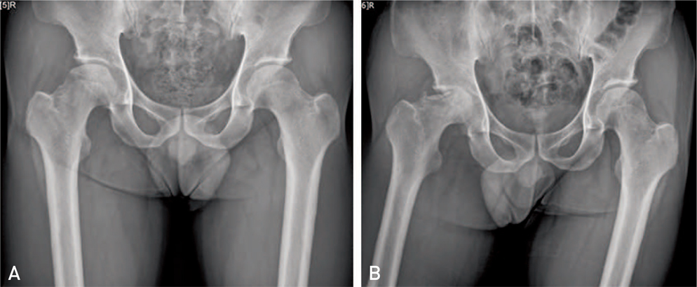
Fig. 1 X-ray findings of rapidly destructive coxarthrosis. (A) Anteroposterior radiograph shows sclerosis of right femoral head. (B) After 4 months, rapid destruction of the right femoral head is noticeable.
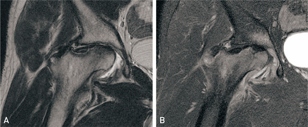
Fig. 2 Magnetic resonance imaging (MRI) findings of rapidly destructive coxarthrosis. (A) Coronal T2-weighted MRI shows severely collapsed femoral head and destroyed acetabulum with extensive bone marrow edema. Intermediate signal intensities within the joint effusion, synovial proliferation, inflammation. (B) Gadolinium-enhanced MRI with fat suppression shows a heterogeneously enhanced synovium, surrounding soft tissue, and bone marrow lesions.
Reference
-
1. Postel M, Kerboull M. Total prosthetic replacement in rapidly destructive arthrosis of the hip joint. Clin Orthop Relat Res. 1970; 72:138–144.2. Kuo A, Ezzet KA, Patil S, Colwell CW Jr. Total hip arthroplasty in rapidly destructive osteoarthritis of the hip: a case series. HSS J. 2009; 5:117–119.
Article3. Ogawa K, Mawatari M, Komine M, et al. Mature and activated osteoclasts exist in the synovium of rapidly destructive coxarthrosis. J Bone Miner Metab. 2007; 25:354–360.
Article4. Lequesne M. [Rapid destructive coxarthritis]. Rhumatologie. 1970; 22:51–63. Dutch.5. Lequesne M. [Rapidly progressing destructive diseases of the hip]. Ann Radiol (Paris). 1993; 36:62–64. French.6. Komiya S, Inoue A, Sasaguri Y, Minamitani K, Morimatsu M. Rapidly destructive arthropathy of the hip. Studies on bone resorptive factors in joint fluid with a theory of pathogenesis. Clin Orthop Relat Res. 1992; (284):273–282.7. Mitrovic DR, Riera H. Synovial, articular cartilage and bone changes in rapidly destructive arthropathy (osteoarthritis) of the hip. Rheumatol Int. 1992; 12:17–22.
Article8. Rosenberg ZS, Shankman S, Steiner GC, Kastenbaum DK, Norman A, Lazansky MG. Rapid destructive osteoarthritis: clinical, radiographic, and pathologic features. Radiology. 1992; 182:213–216.
Article9. Ryu KN, Kim EJ, Yoo MC, Park YK, Sartoris DJ, Resnick D. Ischemic necrosis of the entire femoral head and rapidly destructive hip disease: potential causative relationship. Skeletal Radiol. 1997; 26:143–149.
Article10. Watanabe W, Itoi E, Yamada S. Early MRI findings of rapidly destructive coxarthrosis. Skeletal Radiol. 2002; 31:35–38.
Article11. Charrois O, Kahwaji A, Vastel L, Rosencher N, Courpied JP. Blood loss in total hip arthroplasty for rapidly destructive coxarthrosis. Int Orthop. 2001; 25:22–24.
Article12. Mercuriali F, Inghilleri G. Proposal of an algorithm to help the choice of the best transfusion strategy. Curr Med Res Opin. 1996; 13:465–478.
Article13. Nadler SB, Hidalgo JH, Bloch T. Prediction of blood volume in normal human adults. Surgery. 1962; 51:224–232.14. Boutry N, Paul C, Leroy X, Fredoux D, Migaud H, Cotten A. Rapidly destructive osteoarthritis of the hip: MR imaging findings. AJR Am J Roentgenol. 2002; 179:657–663.
Article15. Lee KH, Sung MS, Kim HM, et al. MR imaging of osteonecrosis of the femoral head with rapidly destructive coxarthrosis. J Korean Orthop Assoc. 2003; 38:105–110.
Article16. Matsumoto F, Uzuki M, Kaneko C, Rikimaru A, Kokubun S, Sawai T. [Expression of matrix metalloproteinases (MMPs) and tissue inhibitor of metalloproteinases (TIMPs) in joint tissues of rapidly destructive coxarthropathy (RDC), analyzed by immunohistochemical study]. Ryumachi. 1997; 37:688–695. Japanese.17. Nam WD, Kim IY, Rhyu KH. Blood loss and transfusion in primary total hip arthroplasty. J Korean Hip Soc. 2006; 18:1–5.
Article
- Full Text Links
-
- Actions
-
Cited
- CITED
-
- Close
- Share
-
- Similar articles
-
- Rapidly Destructive Coxarthrosis in Patients with Rheumatoid Arthritis: Report on 3 Cases
- MR Imaging of Osteonecrosis of the Femoral Head with Rapidly Destructive Coxarthrosis
- Rapidly Destructive Coxarthrosis
- Sequential Bilateral Rapid Destructive Inflammatory Coxarthrosis in a Patient with Human Immunodeficiency Virus
- Total Hip Replacement Arthroplasty in Patient with Idiopathic Thrombocytopenic Purpura: Clinical Experience of Perioperative Management
