Comparison of Bioabsorbable Suture Anchor Fixation on the Tibial Side for Anterior Cruciate Ligament Reconstruction Using Free Soft Tissue Graft: Experimental Laboratory Study on Porcine Bone
- Affiliations
-
- 1Department of Orthopedic Surgery, College of Medicine, Inha University, Incheon, Korea. m9kim@inha.ac.kr
- KMID: 2068688
- DOI: http://doi.org/10.3349/ymj.2014.55.3.760
Abstract
- PURPOSE
The use of graft tissue fixation using bioabsorbable interference screws (BISs) in anterior cruciate ligament (ACL) reconstruction offers various advantages, but limited pullout strength. Therefore, additional tibial fixation is essential for aggressive rehabilitation. We hypothesized that additional graft tissue fixation using bioabsorbable suture anchors (BSA) would provide sufficient pull-out strength.
MATERIALS AND METHODS
Twenty four fresh frozen porcine distal femur and patellar tendon preparations were used. All specimens were divided into three groups based on additional fixation methods: A, isolated BIS; B, BIS and BSA; and C, BIS and post cortical screw. Tensile testing was carried out under an axial load. Ultimate failure load and ultimate failure load after cyclic loading were recorded.
RESULTS
The ultimate failure loads after load to failure testing were 166.8 N in group A, 536.4 N in group B, and 438 N in group C; meanwhile, the ultimate failure loads after load to failure testing with cyclic loading were 140 N in group A, 466.5 N in group B, and 400 N in group C. Stiffness after load to failure testing was 16.5 N/mm in group A, 33.5 N/mm in group B, and 40 N/mm in group C. An additional BSA fixation resulted in a significantly higher ultimate failure load and stiffness than isolated BIS fixation, similar to post screw fixation.
CONCLUSION
Additional fixation using a BSA provided sufficient pullout strength for ACL reconstruction. The ultimate failure load of the BSA technique was similar to that of post cortical screws.
Figure
-

Fig. 1 Graft preparation using Krackow whip stitches, forming four loops at each end of the tendon.

Fig. 2 Additional fixation devices used for this study. (A) 9×25 mm BioScrew®. (B) 6×16.5 mm Duet™. (C) 3.5 mm cortical screw.

Fig. 3 (A) Bioabsorbable suture anchor (Duet™). (B) Separated suture anchor tip and FiberWire® (Arthrex) connecting the tendon. (C) Reassembled bioabsorbable suture anchor.
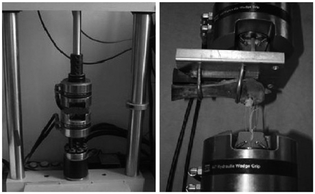
Fig. 4 The 858 Table Top System® testing apparatus is shown with a porcine femur and graft. The free end of the patellar tendon graft was fixed to the inferior clamp of the machine with FiberWire®.
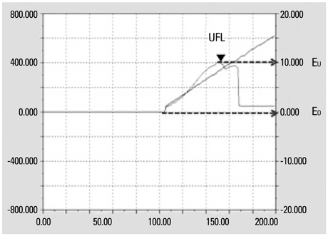
Fig. 5 Load elongation curve of the load to failure test (Elongation=EU-E0). UFL, ultimate failure load.
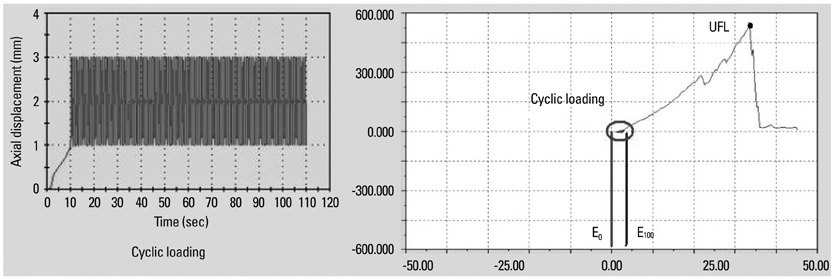
Fig. 6 Load elongation curve of the cyclic loading test (Elongation=E100-E0). UFL, ultimate failure load.
Cited by 1 articles
-
Functional Outcome of Bioabsorbable Suture Anchor and Metal Screw Fixation on Tibial Side for Anterior Cruciate Ligament Reconstruction
Myung-Ku Kim, Ju-Yong Park, Chi-Hoon Ahn
Korean J Sports Med. 2014;32(2):92-96. doi: 10.5763/kjsm.2014.32.2.92.
Reference
-
1. Dragoo JL, Braun HJ, Durham JL, Chen MR, Harris AH. Incidence and risk factors for injuries to the anterior cruciate ligament in National Collegiate Athletic Association football: data from the 2004-2005 through 2008-2009 National Collegiate Athletic Association Injury Surveillance System. Am J Sports Med. 2012; 40:990–995.
Article2. Almqvist KF, Willaert P, De Brabandere S, Criel K, Verdonk R. A long-term study of anterior cruciate ligament allograft reconstruction. Knee Surg Sports Traumatol Arthrosc. 2009; 17:818–822.
Article3. Brand JC Jr, Nyland J, Caborn DN, Johnson DL. Soft-tissue interference fixation: bioabsorbable screw versus metal screw. Arthroscopy. 2005; 21:911–916.
Article4. Giurea M, Zorilla P, Amis AA, Aichroth P. Comparative pull-out and cyclic-loading strength tests of anchorage of hamstring tendon grafts in anterior cruciate ligament reconstruction. Am J Sports Med. 1999; 27:621–625.
Article5. Magen HE, Howell SM, Hull ML. Structural properties of six tibial fixation methods for anterior cruciate ligament soft tissue grafts. Am J Sports Med. 1999; 27:35–43.
Article6. Tetsumura S, Fujita A, Nakajima M, Abe M. Biomechanical comparison of different fixation methods on the tibial side in anterior cruciate ligament reconstruction: a biomechanical study in porcine tibial bone. J Orthop Sci. 2006; 11:278–282.
Article7. Yoo JC, Ahn JH, Kim JH, Kim BK, Choi KW, Bae TS, et al. Biomechanical testing of hybrid hamstring graft tibial fixation in anterior cruciate ligament reconstruction. Knee. 2006; 13:455–459.
Article8. Hill PF, Russell VJ, Salmon LJ, Pinczewski LA. The influence of supplementary tibial fixation on laxity measurements after anterior cruciate ligament reconstruction with hamstring tendons in female patients. Am J Sports Med. 2005; 33:94–101.
Article9. Jansson KA, Linko E, Sandelin J, Harilainen A. A prospective randomized study of patellar versus hamstring tendon autografts for anterior cruciate ligament reconstruction. Am J Sports Med. 2003; 31:12–18.
Article10. Beynnon BD, Johnson RJ, Abate JA, Fleming BC, Nichols CE. Treatment of anterior cruciate ligament injuries, part 2. Am J Sports Med. 2005; 33:1751–1767.
Article11. Clark R, Olsen RE, Larson BJ, Goble EM, Farrer RP. Cross-pin femoral fixation: a new technique for hamstring anterior cruciate ligament reconstruction of the knee. Arthroscopy. 1998; 14:258–267.
Article12. Howell SM, Deutsch ML. Comparison of endoscopic and two-incision techniques for reconstructing a torn anterior cruciate ligament using hamstring tendons. Arthroscopy. 1999; 15:594–606.
Article13. Scheffler SU, Südkamp NP, Göckenjan A, Hoffmann RF, Weiler A. Biomechanical comparison of hamstring and patellar tendon graft anterior cruciate ligament reconstruction techniques: the impact of fixation level and fixation method under cyclic loading. Arthroscopy. 2002; 18:304–315.
Article14. Ma CB, Francis K, Towers J, Irrgang J, Fu FH, Harner CH. Hamstring anterior cruciate ligament reconstruction: a comparison of bioabsorbable interference screw and endobutton-post fixation. Arthroscopy. 2004; 20:122–128.
Article15. Bushnell BD, Byram IR, Weinhold PS, Creighton RA. The use of suture anchors in repair of the ruptured patellar tendon: a biomechanical study. Am J Sports Med. 2006; 34:1492–1499.
Article16. Benedetto KP, Fellinger M, Lim TE, Passler JM, Schoen JL, Willems WJ. A new bioabsorbable interference screw: preliminary results of a prospective, multicenter, randomized clinical trial. Arthroscopy. 2000; 16:41–48.
Article17. Sassmannshausen G, Carr CF. Transcutaneous migration of a tibial bioabsorbable interference screw after anterior cruciate ligament reconstruction. Arthroscopy. 2003; 19:E133–E136.
Article18. Weiler A, Richter M, Schmidmaier G, Kandziora F, Südkamp NP. The EndoPearl device increases fixation strength and eliminates construct slippage of hamstring tendon grafts with interference screw fixation. Arthroscopy. 2001; 17:353–359.
Article19. Suggs J, Wang C, Li G. The effect of graft stiffness on knee joint biomechanics after ACL reconstruction--a 3D computational simulation. Clin Biomech (Bristol, Avon). 2003; 18:35–43.
Article20. Nurmi JT, Sievänen H, Kannus P, Järvinen M, Järvinen TL. Porcine tibia is a poor substitute for human cadaver tibia for evaluating interference screw fixation. Am J Sports Med. 2004; 32:765–771.
Article21. Asahina S, Muneta T, Ishibashi T, Yamamoto H. Effects of knee flexion angle at graft fixation on the outcome of anterior cruciate ligament reconstruction. Arthroscopy. 1996; 12:70–75.
Article22. Leppälä J, Kannus P, Natri A, Pasanen M, Sievänen H, Vuori I, et al. Effect of anterior cruciate ligament injury of the knee on bone mineral density of the spine and affected lower extremity: a prospective one-year follow-Up study. Calcif Tissue Int. 1999; 64:357–363.
Article23. Vuori I, Heinonen A, Sievänen H, Kannus P, Pasanen M, Oja P. Effects of unilateral strength training and detraining on bone mineral density and content in young women: a study of mechanical loading and deloading on human bones. Calcif Tissue Int. 1994; 55:59–67.
Article24. Sim JA, Kwak JH, Yang SH, Lee BK. Comparative biomechanical study of the Ligament Plate and other fixation devices in ACL reconstruction. Int Orthop. 2009; 33:1269–1274.
Article
- Full Text Links
-
- Actions
-
Cited
- CITED
-
- Close
- Share
-
- Similar articles
-
- Functional Outcome of Bioabsorbable Suture Anchor and Metal Screw Fixation on Tibial Side for Anterior Cruciate Ligament Reconstruction
- A Comparison of the Fixation Strengths Provided by Different Intraosseous Tendon Lengths during Anterior Cruciate Ligament Reconstruction: A Biomechanical Study in a Porcine Tibial Model
- Tibial Tunnel Cyst Formation after Anterior Cruciate Ligament Reconstruction Using a Non-Bioabsorbable Interference Screw
- Comparison of Bone Tunnel Widening after ACL Reconstruction between Bioabsorbable Interference Screw and Metallic Interference Screw Fixation
- Optimal Parameters for Sutures Tied to a Post during Anterior Cruciate Ligament Reconstruction: Thread Numbers, Knot Numbers, Suture Techniques and Stitch Numbers: An Experimental Laboratory Study Using Porcine Tendon
