J Korean Neurosurg Soc.
2012 May;51(5):312-315. 10.3340/jkns.2012.51.5.312.
Magnetic Resonance Findings in Two Episodes of Repeated Cerebral Fat Embolisms in a Patient with Autologous Fat Injection into the Face
- Affiliations
-
- 1Department of Radiology, Graduate School of Medicine, Kyung Hee University, Seoul, Korea.
- 2Department of Radiology, Kyung Hee University Hospital, Seoul, Korea. euijkim@hanmail.net
- 3Department of Radiology, Kyung Hee University Hospital at Gangdong, Seoul, Korea.
- 4Department of Neurology, Kyung Hee University Hospital, Seoul, Korea.
- KMID: 2066934
- DOI: http://doi.org/10.3340/jkns.2012.51.5.312
Abstract
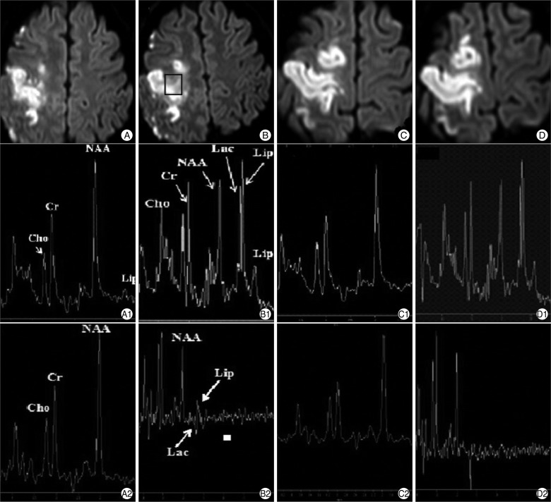
- We report magnetic resonance image (MRI) and magnetic resonance spectroscopy (MRS) findings in a patient of cerebral fat embolism (CFE) occurred in a 26-year-old woman after an autologous fat injection into the face. After initial neurologic symptom onset, MRI and MRS data were obtained two times to investigate repeated CFE. We obtained the MRS data in the two different time intervals and two different echo times to compare the lesions with normal brain parenchyma. The results of MRS data showed that a decrease in N-acetyl-aspartate, an increase in lactate and a very high early peak of free lipids between 0.9 and 1.4 ppm were obtained at the acute infarcted lesion as compared with normal brain parenchyma. In addition, these findings were more clearly detected on short echo time spectrum rather than long spectrum. A close relationship between the clinical manifestations and MRI and MRS findings of the brain can helpful to distinguish CFE with other conditions and to evaluate the cause materials of infarctions rather than conventional MRI or diffusion-weighted imaging.
Keyword
MeSH Terms
Figure
Cited by 1 articles
-
Fat embolism syndrome: a review in cosmetic surgery
Hongil Kim, Bommie Florence Seo, Gregory Randolph Dean Evans
Kosin Med J. 2024;39(3):169-178. doi: 10.7180/kmj.24.126.
Reference
-
1. Egido JA, Arroyo R, Marcos A, Jiménez-Alfaro I. Middle cerebral artery embolism and unilateral visual loss after autologous fat injection into the glabellar area. Stroke. 1993; 24:615–616. PMID: 8465374.
Article2. Guillevin R, Vallée JN, Demeret S, Sonneville R, Bolgert F, Mont'alverne F, et al. Cerebral fat embolism : usefulness of magnetic resonance spectroscopy. Ann Neurol. 2005; 57:434–439. PMID: 15732115.3. Han SJ, Kim JS, Chung SR, Shim YS, Lee KS, Kim YI, et al. Cerebral and ocular fat embolism after autologous fat injection into the face : confirmed by magnetic resonance spectroscopy. J Korean Neurol Assoc. 2006; 24:399–401.4. Kim HJ, Lee CH, Lee SH, Moon TY. Magnetic resonance imaging and histologic findings of experimental cerebral fat embolism. Invest Radiol. 2003; 38:625–634. PMID: 14501490.
Article5. Lee YS, Park SH, Hamm IS. Cerebral fat embolism with multiple rib and thoracic spinal fractures. J Korean Neurosurg Soc. 2004; 36:408–411.6. Robinson EF, Soloway HB. Experimental cerebral fat embolism. Distribution of radioactive triolein following internal carotid introduction. Arch Neurol. 1971; 24:419–422. PMID: 5554865.
- Full Text Links
- Actions
-
Cited
- CITED
-
- Close
- Share
- Similar articles
-
- A Case of Acute Stroke after Autologous Fat Injection
- Cerebral and Ocular Fat Embolism after Autologous Fat Injection into the Face: Confirmed by Magnetic Resonance Spectroscopy
- Ophthalmic Artery Obstruction and Cerebral Infarction Following Periocular Injection of Autologous Fat
- Autologous fat injection and collagen impantation for the correction of facial atroply
- The Simple Correction of Deep Nasolabial Fold by Autologous Microfat Fat Injection


