Obstet Gynecol Sci.
2015 Sep;58(5):405-408. 10.5468/ogs.2015.58.5.405.
Prenatal diagnosis of congenital mesoblastic nephroma
- Affiliations
-
- 1Department of Obstetrics and Gynecology, Samsung Medical Center, Sungkyunkwan University School of Medicine, Seoul, Korea. ohsymd@skku.edu
- 2Department of Pathology, Samsung Medical Center, Sungkyunkwan University School of Medicine, Seoul, Korea.
- KMID: 2058466
- DOI: http://doi.org/10.5468/ogs.2015.58.5.405
Abstract
- Congenital mesoblastic nephroma is a rare renal tumor that is diagnosed during pregnancy and is associated with polyhydramnios, prematurity, and neonatal hypertension. Differential diagnoses include Wilms tumor, adrenal neuroblastoma, and other abdominal tumors. We report a case of congenital mesoblastic nephroma detected by prenatal ultrasonography as a large fetal renal mass with polyhydramnios at 32 weeks of gestation. Ultrasonography showed a 6x6-cm complex, solid, hyperechoic, round mass in the right kidney. At 35 weeks of gestation, the patient was admitted with preterm premature rupture of membranes and the baby was delivered vaginally. Postnatal ultrasonography and computed tomography showed a heterogeneous solid mass on the right kidney. At the end of the first week of life, a right nephrectomy was performed and subsequent pathological examination confirmed a cellular variant of congenital mesoblastic nephroma with a high mitotic count. Postoperative adjuvant chemotherapy was administered. The newborn was discharged in good condition.
MeSH Terms
Figure
-
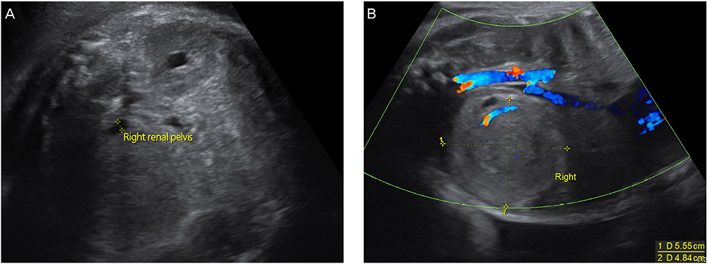
Fig. 1 Prenatal ultrasonography reveals a well-circumscribed, solid, hyperechoic mass in the right kidney, measuring 5.6×4.8 cm. (A) Transverse section. (B) Longitudinal section.
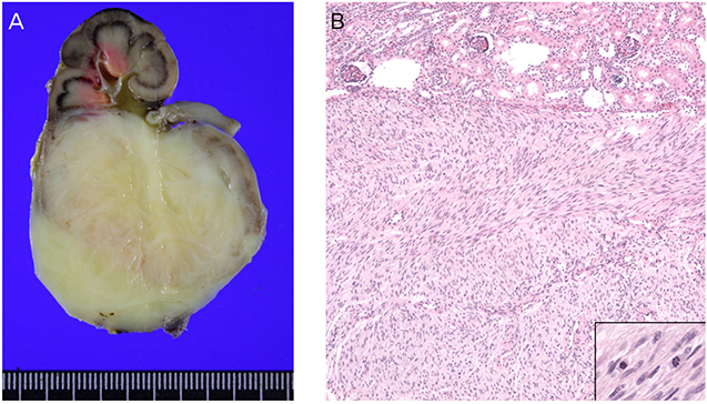
Fig. 2 Nephrectomy: cut sections and pathologic findings. (A) The mass originated from the lower pole of the right kidney. The cut surface is yellowish white with a whirling pattern. Lower, mass; upper, normal. (B) Hematoxylin and eosin stain, ×100. Microscopically, the mass is composed of intersecting bundles of spindle cells with frequent mitoses (inset).
Reference
-
1. Chen WY, Lin CN, Chao CS, Yan-Sheng Lin M, Mak CW, Chuang SS, et al. Prenatal diagnosis of congenital mesoblastic nephroma in mid-second trimester by sonography and magnetic resonance imaging. Prenat Diagn. 2003; 23:927–931.2. Glick RD, Hicks MJ, Nuchtern JG, Wesson DE, Olutoye OO, Cass DL. Renal tumors in infants less than 6 months of age. J Pediatr Surg. 2004; 39:522–525.3. Shapiro E. Upper urinary tract anomalies and perinatal renal tumors. Clin Perinatol. 2014; 41:679–694.4. Powis M. Neonatal renal tumours. Early Hum Dev. 2010; 86:607–612.5. Miller OF, Kolon TF. Hyperreninemia and congenital mesoblastic nephroma: case report and review of the literature. Urology. 2000; 55:775.6. Ko SM, Kim MJ, Im YJ, Park KI, Lee MJ. Cellular mesoblastic nephroma with liver metastasis in a neonate: prenatal and postnatal diffusion-weighted MR imaging. Korean J Radiol. 2013; 14:361–365.7. Garnier S, Maillet O, Haouy S, Saguintaah M, Serre I, Galifer RB, et al. Prenatal intrarenal neuroblastoma mimicking a mesoblastic nephroma: a case report. J Pediatr Surg. 2012; 47:e21–e23.8. Rubenstein SC, Benacerraf BR, Retik AB, Mandell J. Fetal suprarenal masses: sonographic appearance and differential diagnosis. Ultrasound Obstet Gynecol. 1995; 5:164–167.9. Wang ZP, Li K, Dong KR, Xiao XM, Zheng S. Congenital mesoblastic nephroma: clinical analysis of eight cases and a review of the literature. Oncol Lett. 2014; 8:2007–2011.10. Woodward PJ, Sohaey R, Kennedy A, Koeller KK. From the archives of the AFIP: a comprehensive review of fetal tumors with pathologic correlation. Radiographics. 2005; 25:215–242.11. Shibahara H, Mitsuo M, Fujimoto K, Muranaka J, Sawai H, Bessho T, et al. Prenatal sonographic diagnosis of a fetal renal mesoblastic nephroma occurring after transfer of a cryopreserved embryo. Hum Reprod. 1999; 14:1324–1327.12. Kim CH, Kim YH, Cho MK, Kim KM, Ha JA, Joo EH, et al. A case of fetal congenital mesoblastic nephroma with oligohydramnios. J Korean Med Sci. 2007; 22:357–361.13. Vujanic GM, Delemarre JF, Moeslichan S, Lam J, Harms D, Sandstedt B, et al. Mesoblastic nephroma metastatic to the lungs and heart: another face of this peculiar lesion: case report and review of the literature. Pediatr Pathol. 1993; 13:143–153.
