A new bite block for panoramic radiographs of anterior edentulous patients: A technical report
- Affiliations
-
- 1Department of Oral and Maxillofacial Radiology and Dental Research Institute, School of Dentistry, Seoul National University, Seoul, Korea. hmslsh@snu.ac.kr
- 2Division of Oral and Maxillofacial Radiology, Department of Basic Science, Faculty of Dentistry, University of Health Sciences, Vientiane, Laos.
- KMID: 2054214
- DOI: http://doi.org/10.5624/isd.2015.45.2.117
Abstract
- PURPOSE
Panoramic radiographs taken using conventional chin-support devices have often presented problems with positioning accuracy and reproducibility. The aim of this report was to propose a new bite block for panoramic radiographs of anterior edentulous patients that better addresses these two issues.
MATERIALS AND METHODS
A new panoramic radiography bite block similar to the bite block for dentulous patients was developed to enable proper positioning stability for edentulous patients. The new bite block was designed and implemented in light of previous studies. The height of the new bite block was 18 mm and to compensate for the horizontal edentulous space, its horizontal width was 7 mm. The panoramic radiographs using the new bite block were compared with those using the conventional chin-support device.
RESULTS
Panoramic radiographs taken with the new bite block showed better stability and bilateral symmetry than those taken with the conventional chin-support device. Patients also showed less movement and more stable positioning during panoramic radiography with the new bite block.
CONCLUSION
Conventional errors in panoramic radiographs of edentulous patients could be caused by unreliability of the chin-support device. The newly proposed bite block for panoramic radiographs of edentulous patients showed better reliability. Further study is required to evaluate the image quality and reproducibility of images with the new bite block.
Figure
-
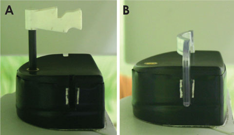
Fig. 1 A. The conventional bite block for anterior edentulous patients. B. The chin-support device for anterior edentulous patients.
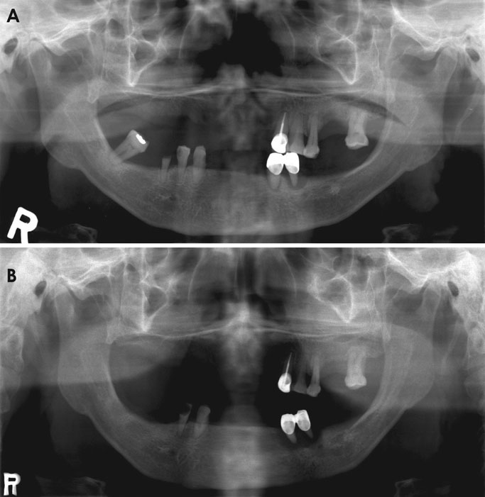
Fig. 2 A and B. Two panoramic radiographs acquired from the same anterior edentulous patient using a conventional chin-support device show lack of reproducibility in the inter-maxillary vertical dimension.
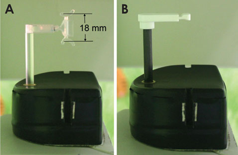
Fig. 3 A. The new bite block for anterior edentulous patients. B. The conventional standard bite block for anterior dentulous patients.

Fig. 4 A. The calculation to determine the height of the new bite block. B. The horizontal distance calculation to determine the antero-posterior dimension of the new bite block.
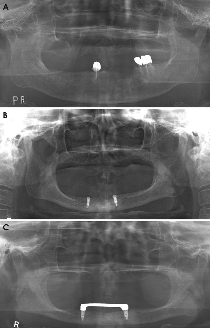
Fig. 5 panoramic radiographs acquired from anterior edentulous patients using the conventional chin-support device. A. The radiograph shows insufficient inter-maxillary vertical height. B. The radiograph shows asymmetry although the patient's jaw is not asymmetrical. C. The panoramic radiographs acquired from an anterior edentulous patient using the conventional chin-support device show horizontal magnification.
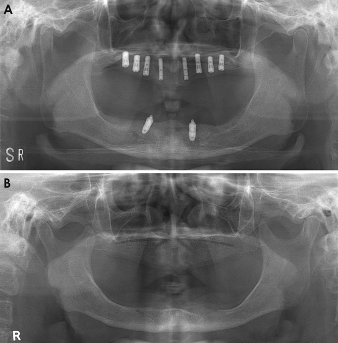
Fig. 6 A and B. The panoramic radiographs taken using the new bite block showed a more suitable vertical height and antero-posterior position, and better bilateral symmetry than the panoramic radiographs taken using the chin-support device.
Reference
-
1. Dhillon M, Raju SM, Verma S, Tomar D, Mohan RS, Lakhanpal M, et al. Positioning errors and quality assessment in panoramic radiography. Imaging Sci Dent. 2012; 42:207–212.
Article2. Glass BJ, Seals RR Jr, Williams EO. Common errors in panoramic radiography of edentulous patients. J Prosthodont. 1994; 3:68–73.
Article3. Batenburg RH, Stellingsma K, Raghoebar GM, Vissink A. Bone height measurements on panoramic radiographs: the effect of shape and position of edentulous mandibles. Oral Surg Oral Med Oral Pathol Oral Radiol Endod. 1997; 84:430–435.4. Kogon S, Bohay R, Stephens R. A survey of the radiographic practices of general dentists for edentulous patients. Oral Surg Oral Med Oral Pathol Oral Radiol Endod. 1995; 80:365–368.
Article5. Spyropoulos ND, Patsakas AJ, Angelopoulos AP. Findings from radiographs of the jaws of edentulous patients. Oral Surg Oral Med Oral Pathol. 1981; 52:455–459.
Article6. Swenson HM, Hudson JR. Roentgenographic examination of edentulous patients. J Prosthet Dent. 1967; 18:304–307.
Article7. Ortman LF, Hausmann E, Dunford RG. Skeletal osteopenia and residual ridge resorption. J Prosthet Dent. 1989; 61:321–325.
Article8. Soikkonen K, Ainamo A, Xie Q. Height of the residual ridge and radiographic appearance of bony structure in the jaws of clinically edentulous elderly people. J Oral Rehabil. 1996; 23:470–475.
Article9. Wical KE, Swoope CC. Studies of residual ridge resorption. I. Use of panoramic radiographs for evaluation and classification of mandibular resorption. J Prosthet Dent. 1974; 32:7–12.10. Kaffe I, Ardekian L, Gelerenter I, Taicher S. Location of the mandibular foramen in panoramic radiographs. Oral Surg Oral Med Oral Pathol. 1994; 78:662–669.
Article11. Scarfe WC, Eraso FE, Farman AG. Characteristics of the Orthopantomograph OP 100. Dentomaxillofac Radiol. 1998; 27:51–57.
Article12. Park JW, Huh KH, Yi WJ, Heo MS, Lee SS, Choi SC. Comparison of the reproducibility of panoramic radiographs between dentulous and edentulous patients. Imaging Sci Dent. 2014; 44:95–102.
Article13. Volchansky A, Cleaton-Jones P. Clinical crown height (length) - a review of published measurements. J Clin Periodontol. 2001; 28:1085–1090.
Article14. Tong H, Kwon D, Shi J, Sakai N, Enciso R, Sameshima GT. Mesiodistal angulation and faciolingual inclination of each whole tooth in 3-dimensional space in patients with near-normal occlusion. Am J Orthod Dentofacial Orthop. 2012; 141:604–617.
Article15. Sato M, Motoyoshi M, Hirabayashi M, Hosoi K, Mitsui N, Shimizu N. Inclination of the occlusal plane is associated with the direction of the masticatory movement path. Eur J Orthod. 2007; 29:21–25.
Article16. Canger EM, Celenk P. Radiographic evaluation of alveolar ridge heights of dentate and edentulous patients. Gerodontology. 2012; 29:17–23.
Article
- Full Text Links
-
- Actions
-
Cited
- CITED
-
- Close
- Share
-
- Similar articles
-
- Comparison of the reproducibility of panoramic radiographs between dentulous and edentulous patients
- Complete denture fabrication of a skeletal class III edentulous patient considering anterior neutral zone: a case report
- Screening for variations in anterior digastric musculature prior to correction of post-traumatic anterior open bite by injection of botulinum toxin type A: a technical note
- Residual bone height measured by panoramic radiography in older edentulous Korean patients
- Prediction of osteoporosis using fractal analysis on periapical and panoramic radiographs
