Can pterygoid plate asymmetry be linked to temporomandibular joint disorders?
- Affiliations
-
- 1OIC, OMFS IMPATH Research Group, Department of Imaging and Pathology, Faculty of Medicine, University of Leuven and Oral and Maxillofacial Surgery, University Hospitals Leuven, Belgium. reinhilde.jacobs@uzleuven.be
- 2Oral and Maxillofacial Radiology, Post-Graduate School, Universidad Privada Cayetano Heredia, Peru.
- 3Stomatology and Maxillofacial Surgery, Department of Oral Health Sciences, KU Leuven, Belgium.
- KMID: 2054210
- DOI: http://doi.org/10.5624/isd.2015.45.2.89
Abstract
- PURPOSE
This study was performed to evaluate the relationship between pterygoid plate asymmetry and temporomandibular joint disorders.
MATERIALS AND METHODS
Cone-beam computed tomography (CBCT) images of 60 patients with temporomandibular disorders (TMD) involving pain were analyzed and compared with images of 60 age- and gender-matched controls. Three observers performed linear measurements of the lateral pterygoid plates.
RESULTS
Statistically significant differences were found between measurements of the lateral pterygoid plates on the site that had pain and the contralateral site (p<0.05). The average length of the lateral pterygoid plates (LPPs) in patients with TMD was 17.01+/-3.64 mm on the right side and 16.21+/-3.51 mm on the left side, and in patients without TMD, it was 11.86+/-1.97 mm on the right side and 11.98+/-1.85 mm on the left side. Statistically significant differences in the LPP length, measured on CBCT, were found between patients with and without TMD (p<0.05). The inter-examiner reliability obtained in this study was very high for all the examiners (0.99, 95% confidence interval: 0.98-0.99).
CONCLUSION
Within the limits of the present study, CBCT lateral pterygoid plate measurements at the side with TMD were found to be significantly different from those on the side without TMD. More research is needed to explore potential etiological correlations and implications for treatment.
Keyword
MeSH Terms
Figure
-
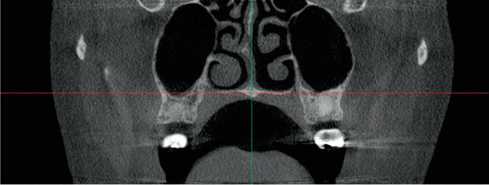
Fig. 1 Coronal cone-beam computed tomography (CT) image shows the intersection of the midsagittal plane with the nasal cavity floor plane.
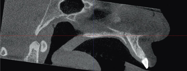
Fig. 2 Sagittal cone-beam CT image shows the midsagittal plane and the long axis of the palatal plane selected in order to obtain a proper axial view of the outer wings of the bilateral pterygoid tubercle.
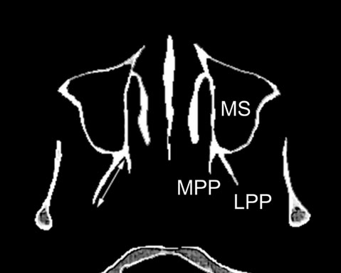
Fig. 3 Axial cone-beam CT image shows the measurement in the lateral pterygoid plate (LPP), medial pterygoid plate (MPP), and maxillary sinus (MS).
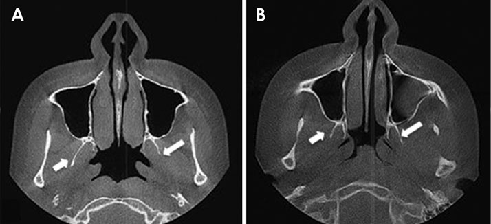
Fig. 4 A. Axial cone-beam CT image shows the differences between the left and the right pterygoid plates of a 22-year-old man with temporomandibular disorder (TMD) B. Axial cone-beam CT image shows no differences between the left and the right pterygoid plates on a matched control.
Reference
-
1. Okeson JP. Current terminology and diagnostic classification schemes. Oral Surg Oral Med Oral Pathol Oral Radiol Endod. 1997; 83:61–64.
Article2. Murray GM, Bhutada M, Peck CC, Phanachet I, Sae-Lee D, Whittle T. The human lateral pterygoid muscle. Arch Oral Biol. 2007; 52:377–380.
Article3. Fujita S, Iizuka T, Dauber W. Variation of heads of lateral pterygoid muscle and morphology of articular disc of human temporomandibular joint - anatomical and histological analysis. J Oral Rehabil. 2001; 28:560–571.
Article4. Liu ZJ, Wang HY, Pu WY. A comparative electromyographic study of the lateral pterygoid muscle and arthrography in patients with temporomandibular joint disturbance syndrome sounds. J Prosthet Dent. 1989; 62:229–233.5. Grunheid T, Langenbach GE, Korfage JA, Zentner A, van Eijden TM. The adaptive response of jaw muscles to varying functional demands. Eur J Orthod. 2009; 31:596–612.
Article6. Osborn AG. Radiology of the pterygoid plates and pterygopalatine fossa. AJR Am J Roentgenol. 1979; 132:389–394.
Article7. List T, Dworkin SF. Comparing TMD diagnoses and clinical findings at Swedish and US TMD centers using research diagnostic criteria for temporomandibular disorders. J Orofac Pain. 1996; 10:240–253.8. Greene CS. The etiology of temporomandibular disorders: implications for treatment. J Orofac Pain. 2001; 15:93–116.9. Schwartz LL. Pain associated with the temporomandibular joint. J Am Dent Assoc. 1955; 51:394–397.
Article10. Schwartz M, Freund B. Treatment of temporomandibular disorders with botulinum toxin. Clin J Pain. 2002; 18:S198–S203.
Article11. Freund B, Schwartz M, Symington JM. Botulinum toxin: new treatment for temporomandibular disorders. Br J Oral Maxillofac Surg. 2000; 38:466–471.
Article12. Bakke M, Moller E, Werdelin LM, Dalager T, Kitai N, Kreiborg S. Treatment of severe temporomandibular joint clicking with botulinum toxin in the lateral pterygoid muscle in two cases of anterior disc displacement. Oral Surg Oral Med Oral Pathol Oral Radiol Endod. 2005; 100:693–700.
Article13. Bakathir AA, Margasahayam MV, Al-Ismaily MI. Maxillary hyperplasia and hyperostosis cranialis: a rare manifestation of renal osteodystrophy in a patient with hyperparathyroidism secondary to chronic renal failure. Saudi Med J. 2008; 29:1815–1818.14. Sharma R. Pterygoid disjunction for internal derangement of temporomandibular joint. J Maxillofac Oral Surg. 2011; 10:142–147.
Article15. Kannan SV. Pterygoid dysjunction: new minimally invasive technique for the treatment of painful temporomandibular joint dysfunction. J Craniofac Surg. 2010; 21:1264–1269.16. Camilleri AE. Craniofacial fibrous dysplasia. J Laryngol Otol. 1991; 105:662–666.
Article17. Ueki K, Hashiba Y, Marukawa K, Nakagawa K, Okabe K, Yamamoto E. Determining the anatomy of the descending palatine artery and pterygoid plates with computed tomography in Class III patients. J Craniomaxillofac Surg. 2009; 37:469–473.
Article18. Sato S, Kawamura H, Motegi K, Takahashi K. Morphology of the mandibular fossa and the articular eminence in temporomandibular joints with anterior disc displacement. Int J Oral Maxillofac Surg. 1996; 25:236–238.19. Peck CC, Hannam AG. Human jaw and muscle modelling. Arch Oral Biol. 2007; 52:300–304.
Article
- Full Text Links
-
- Actions
-
Cited
- CITED
-
- Close
- Share
-
- Similar articles
-
- A Rare Case of Solitary Osteochondroma at the Temporomandibular Joint: A Case Report
- One-stage total reconstruction of temporomandibular joint ankylosis and facial asymmetry
- The relationship between mandibular asymmetry and temporomandibular joint disc displacement on mri
- A study on simultation of the mandibular movement of the patients with temporomandibular joint disorder
- Unilateral intraoral vertical ramus osteotomy and sagittal split ramus osteotomy for the treatment of asymmetric mandibles
