Tuberc Respir Dis.
2010 Feb;68(2):97-100. 10.4046/trd.2010.68.2.97.
A Case of Pneumocystis Pneumonia Mimicking Acute Eosinophilic Pneumonia in a Patient with AIDS
- Affiliations
-
- 1Department of Internal Medicine, Inje University Seoul Paik Hospital, Inje University College of Medicine, Seoul, Korea. pulho@korea.com
- KMID: 2050592
- DOI: http://doi.org/10.4046/trd.2010.68.2.97
Abstract
- 73-year-old man was admitted with a sudden onset of dyspnea. He had never smoked. The chest radiograph and computed tomography revealed bilateral ground glass opacity and an enlarging perihilar consolidation with lymphadenopathies. There was a higher percentage of eosinophils (72%) in the bronchoalveolar lavage fluid (BALF) than normal. The patient was diagnosed with acute eosinophilic pneumonia and managed with steroid. Pneumocystis pneumonia (PCP) was diagnosed by an examination of the BALF, and the patient was treated with trimethoprim-sulphamethoxazole. The patient tested positive to the HIV antibody and the peripheral blood CD-4 positive lymphocyte count was only 33/microliter. The percentage of eosinophils in the BALF can increase in some cases of PCP that is complicated with AIDS. Only a few cases of eosinophilic pneumonia associated with PCP pneumonia have been reported in patients with AIDS but there are no case reports in Korea. This case highlights the need to consider PCP when the percentage of eosinophils in the BALF is elevated.
MeSH Terms
Figure
-
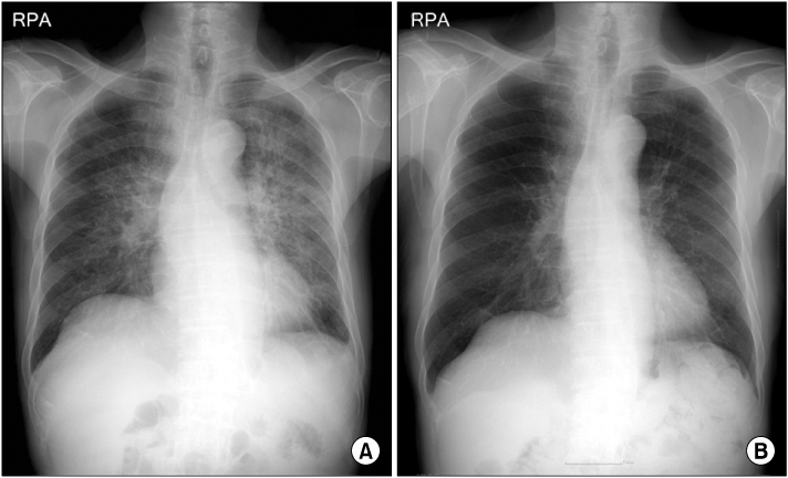
Figure 1 Chest PA at emergency room (A) and 14th hospital day (B). The bilateral hilar enlargement and ground glass opecities with increased interstitial markings were showed and improved 2 weeks later.

Figure 2 Chest computed tomography on admission. The bilateral perihilar ground glass opecities with increased interstitial markings were showed.

Figure 3 Bronchoalveolar lavage fluid. (A) The nucleus of eosinophils contains one to two lobes. The cytoplasm is abundant with a full complement of bright reddish specific granules (Giemsa stain, ×400). (B) Pneumocystis jirovecci aggregates of many discoid shaped cysts with thick, darkly stained, silver-positive capsule and a central tiny dot (GMS stain, ×400).
Reference
-
1. Sadaghdar H, Huang ZB, Eden E. Correlation of bronchoalveolar lavage findings to severity of Pneumocystis carinii pneumonia in AIDS: evidence for the development of high-permeability pulmonary edema. Chest. 1992. 102:63–69.2. Thomas CF Jr, Limper AH. Pneumocystis pneumonia. N Engl J Med. 2004. 350:2487–2498.3. Nuesch R, Bellini C, Zimmerli W. Pneumocystis carinii pneumonia in human immunodeficiency virus (HIV)-positive and HIV-negative immunocompromised patients. Clin Infect Dis. 1999. 29:1519–1523.4. Chung JS, Cho GJ, Kwak IS, Rha HY. A clinical study and prognostic factors for short-term survival of Pneumocystis carinii pneumonia in patients with AIDS. Korean J Med. 1998. 54:488–493.5. Jain P, Sandur S, Meli Y, Arroliga AC, Stoller JK, Mehta AC. Role of flexible bronchoscopy in immunocompromised patients with lung infiltrates. Chest. 2004. 125:712–722.6. Tasaka S, Hasegawa N, Kobayashi S, Yamada W, Nishimura T, Takeuchi T, et al. Serum indicators for the diagnosis of Pneumocystis pneumonia. Chest. 2007. 131:1173–1180.7. Cruciani M, Marcati P, Malena M, Bosco O, Serpelloni G, Mengoli C. Meta-analysis of diagnostic procedures for Pneumocystis carinii pneumonia in HIV-1-infected patients. Eur Respir J. 2002. 20:982–989.8. Smith RL, el-Sadr WM, Lewis M. Correlation of bronchoalveolar lavage cell populations with clinical severity of Pneumocystis carinii pneumonia. Chest. 1988. 93:60–64.9. Itoh M, Nakamura H, Nemoto K, Komiyama M, Hatao H, Shimizudani N, et al. A case of AIDS-complicated lung infection mimicking acute eosinophilic pneumonia. Nihon Kokyuki Gakkai Zasshi. 2006. 44:589–594.10. Jeong YJ, Kim KI, Seo IJ, Lee CH, Lee KN, Kim KN, et al. Eosinophilic lung diseases: a clinical, radiologic, and pathologic overview. Radiographics. 2007. 27:617–637. discussion 637-9.11. Fleury-Feith J, Van Nhieu JT, Picard C, Escudier E, Bernaudin JF. Bronchoalveolar lavage eosinophilia associated with Pneumocystis carinii pneumonitis in AIDS patients: comparative study with non-AIDS patients. Chest. 1989. 95:1198–1201.
- Full Text Links
-
- Actions
-
Cited
- CITED
-
- Close
- Share
-
- Similar articles
-
- A Case of Pneumocystis carinii Pneumonia with Febrile Neutropenia in Acute Lymphoblastic Leukemia
- Thirty six-year-old man presenting acute respiratory failure
- A Clinical Study of 15 Cases of Pneumocystis Carinii Pneumonia
- A Case of Acute eosinophilic pneumonia
- Two Autopsy Cases of Pneumocystis Carinii Pneumonia
