Chonnam Med J.
2012 Dec;48(3):179-182. 10.4068/cmj.2012.48.3.179.
Computed Tomographic Evaluation of Thyroglossal Duct Cysts in Children Under 11 Years of Age
- Affiliations
-
- 1Department of Otolaryngology-Head and Neck Surgery, Chonnam National University Medical School & Chonnam National University Hwasun Hospital, Hwasun, Korea. joonkyoo@jnu.ac.kr
- 2Department of Radiology, Chonnam National University Medical School & Chonnam National University Hwasun Hospital, Hwasun, Korea.
- 3Research Institute of Medical Sciences, Chonnam National University Medical School & Chonnam National University Hwasun Hospital, Hwasun, Korea.
- KMID: 2048809
- DOI: http://doi.org/10.4068/cmj.2012.48.3.179
Abstract
- The purpose of this study was to review the computed tomography (CT) features of thyroglossal duct cysts (TDCs) in children less than 11 years of age. A retrospective chart review was performed at Chonnam National University Hospital for the period of March 2005 to June 2011. CT scans of 16 patients having TDCs were evaluated for the following features: site of the mass, relationship to the midline, walls, margins, internal septa, rim enhancement, internal density, and the presence or absence of the thyroid gland. Of the 16 lesions, 8 (50%) were located in the midline and 12 (75%) were infrahyoid in location. Twelve (75%) of the 16 patients had well-circumscribed walls and peripheral rim enhancement. Internal septa were seen in four of the cysts, and all but one of the cysts demonstrated homogeneous or low-density attenuation. The most common CT findings of TDCs in children less than 11 years of age were a homogeneous or low-density lesion. TDCs in children under the age of 11 years were mostly located in the infrahyoid neck.
Keyword
Figure
-
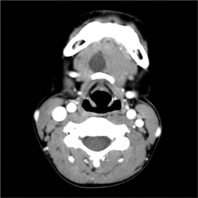
FIG. 1 Thyroglossal duct cyst in a 5-year-old female (case number 1). Axial contrast-enhanced CT scans show a homogeneous and low-density lesion in the anterior midline of the neck.
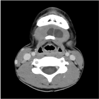
FIG. 2 Thyroglossal duct cyst in a 7-year-old male (case number 8). Axial contrast-enhanced CT scans show a well-circumscribed and septated lesion in the left side of the midline neck.
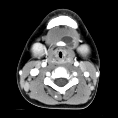
FIG. 3 Thyroglossal duct cyst in a 4-year-old male (case number 14). Axial contrast-enhanced CT scans show a peripheral rim enhancement of the lesion.
Reference
-
1. Allard RH. The thyroglossal cyst. Head Neck Surg. 1982. 5:134–146.
Article2. Davenport M. ABC of general surgery in children. Lumps and swellings of the head and neck. BMJ. 1996. 312:368–371.
Article3. Ahuja AT, Wong KT, King AD, Yuen EH. Imaging for thyroglossal duct cyst: the bare essentials. Clin Radiol. 2005. 60:141–148.
Article4. Reede DL, Bergeron RT, Som PM. CT of thyroglossal duct cysts. Radiology. 1985. 157:121–125.
Article5. Ward RF, Selfe RW, St Louis L, Bowling D. Computed tomography and the thyroglossal duct cyst. Otolaryngol Head Neck Surg. 1986. 95:93–98.
Article6. Friedman ER, John SD. Imaging of pediatric neck masses. Radiol Clin North Am. 2011. 49:617–632.
Article7. Woo EK, Connor SE. Computed tomography and magnetic resonance imaging appearances of cystic lesions in the suprahyoid neck: a pictorial review. Dentomaxillofac Radiol. 2007. 36:451–458.
Article8. Wadsworth DT, Siegel MJ. Thyroglossal duct cysts: variability of sonographic findings. AJR Am J Roentgenol. 1994. 163:1475–1477.
Article9. Ahuja AT, King AD, Metreweli C. Sonographic evaluation of thyroglossal duct cysts in children. Clin Radiol. 2000. 55:770–774.
Article10. Pounds LA. Neck masses of congenital origin. Pediatr Clin North Am. 1981. 28:841–844.
Article11. Byrd SE, Richardson M, Gill G, Lee AM. Computer-tomographic appearance of branchial cleft and thyroglossal duct cysts of the neck. Diagn Imaging. 1983. 52:301–312.12. Koeller KK, Alamo L, Adair CF, Smirniotopoulos JG. Congenital cystic masses of the neck: radiologic-pathologic correlation. Radiographics. 1999. 19:121–146.13. Lin ST, Tseng FY, Hsu CJ, Yeh TH, Chen YS. Thyroglossal duct cyst: a comparison between children and adults. Am J Otolaryngol. 2008. 29:83–87.
Article14. Lee DH, Jung SH, Yoon TM, Lee JK, Joo YE, Lim SC. Preoperative computed tomography of suspected thyroglossal duct cysts in children under 10-years-of-age. Int J Pediatr Otorhinolaryngol. 2012. [Epub ahead of print].
Article15. King AD, Ahuja AT, Mok CO, Metreweli C. MR imaging of thyroglossal duct cysts in adults. Clin Radiol. 1999. 54:304–308.
Article16. Gupta P, Maddalozzo J. Preoperative sonography in presumed thyroglossal duct cysts. Arch Otolaryngol Head Neck Surg. 2001. 127:200–202.
Article17. Huoh KC, Durr ML, Meyer AK, Rosbe KW. Comparison of imaging modalities in pediatric thyroglossal duct cysts. Laryngoscope. 2012. 122:1405–1408.
Article18. Solomon JR, Rangecroft L. Thyroglossal-duct lesions in childhood. J Pediatr Surg. 1984. 19:555–561.
Article19. Marianowski R, Ait Amer JL, Morisseau-Durand MP, Manach Y, Rassi S. Risk factors for thyroglossal duct remnants after Sistrunk procedure in a pediatric population. Int J Pediatr Otorhinolaryngol. 2003. 67:19–23.
Article
