Right sided descending and sigmoid colon: its embryological basis and clinical implications
- Affiliations
-
- 1Department of Anatomy, Lady Hardinge Medical College and SSK Hospital, New Delhi, India. drpreetishrivastava@gmail.com
- KMID: 2046772
- DOI: http://doi.org/10.5115/acb.2013.46.4.299
Abstract
- Anatomical variations of the colon are described by various authors, but the occurrence of right sided descending and sigmoid colon is rare and has not been reported. We found that the anomalous right-sided descending and sigmoid colon had four parts. The proximal segment of the first part consisted of the descending colon extending across the midline from the splenic flexure to the portion supplied by the left colic artery. The distal segment was supplied by the superior sigmoid artery. The second and third parts formed a loop in the right lumbar region anterior to the lumbar cecum. The fourth part was in the lesser pelvis, extending from right sacroiliac joint to the third sacral body. Parts two, three, and four were supplied by the inferior sigmoid artery, which arose from the right side of the inferior mesenteric artery. The ascending and transverse colon was normally placed. This is a rare anomaly that has not been reported so far in adults and is of immense importance to interventional radiologists and colorectal surgeons. The embryological basis of such an anomaly is defective fixation occurring as early as the 12th-17th week of intrauterine life.
Keyword
MeSH Terms
Figure
-

Fig. 1 Anterior view of anomalous colon in situ after removing jejunum and ileum. 1, part 1 of anomalous colon; 2, part 2 of anomalous colon; 3, part 3 of anomalous colon; 4, part 4 of anomalous colon; S, superior; I, inferior; L, left; R, right; a, hepatic flexure; b, ascending colon; c, sigmoid loop anterior to caecum; d, right iliopsoas muscle; e, transverse mesocolon; f, transverse colon; g, superior mesenteric vessels; h, abdominal aorta; i, cut end of ileo-cecal junction.

Fig. 2 Photograph showing the location of the right-sided sigmoid loop. a, ileum; b, sigmoid loop; c, jejunum; d, duodeno-jejunal junction; e, splenic flexure; f, common mesentery.
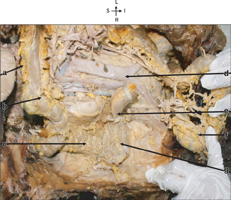
Fig. 3 Fig showing complete large intestine after removal of small intestine. The sigmoid loop has been displaced downwards and to the left showing its posterior aspect. S, superior; I, inferior; L, left; R, right; a, transverse mesocolon; b, transverse colon; c, ascending colon; d, aorta; e, ileo-caecal junction; f, sigmoid loop turned downwards & to left; g, caecum.
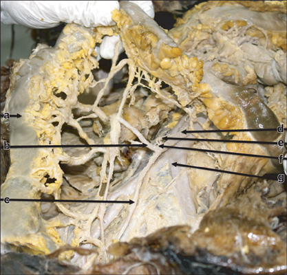
Fig. 4 Photograph showing the vascular supply of the right-sided sigmoid loop. a, sigmoid loop; b, inferior sigmoid artery; c, superior rectal artery; d, inferior mensenteric artery; e, left colic artery; f, superior sigmoid artery; g, bifurcation of aorta.
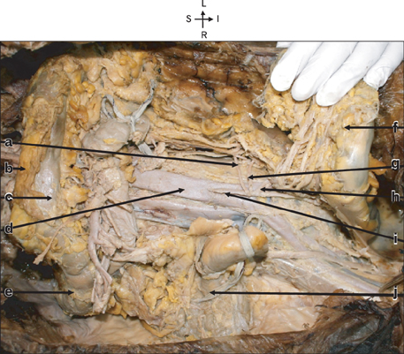
Fig. 5 Photograph showing the branches of inferior mesenteric artery lying posterior to the sigmoid loop which is turned downwards and to the left to expose the artery. S, superior; I, inferior; L, left; R, right; a, left colic artery; b, transverse mesocolon; c, transverse colon; d, aorta; e, hepatic flexure; f, sigmoid loop turned downwards and to the left; g, left sigmoid artery; h, right sigmoid artery; i, inferior mesenteric artery; j, ileo-colic junction.
Reference
-
1. Berardi RS. Anomalies of midgut rotation in the adult. Surg Gynecol Obstet. 1980; 151:113–124.2. Komiyama M, Shimada Y. A case of a right-sided sigmoid colon. Kaibogaku Zasshi. 1991; 66:537–540.3. Madiba TE, Haffajee MR. Sigmoid colon morphology in the population groups of Durban, South Africa, with special reference to sigmoid volvulus. Clin Anat. 2011; 24:441–453.4. Madiba TE, Haffajee MR. Anatomical variations in the level of origin of the sigmoid colon from the descending colon and the attachment of the sigmoid mesocolon. Clin Anat. 2010; 23:179–185.5. Fiorella DJ, Donnelly LF. Frequency of right lower quadrant position of the sigmoid colon in infants and young children. Radiology. 2001; 219:91–94.6. Saxena AK, Sodhi KS, Tirumani S, Mumtaz HA, Narasimha Rao KL, Khandelwal N. Position of a sigmoid colon in right iliac fossa in children: a retrospective study. J Indian Assoc Pediatr Surg. 2011; 16:93–96.7. Hadar H, Gadoth N. Positional relations of colon and kidney determined by perirenal fat. AJR Am J Roentgenol. 1984; 143:773–776.8. Faure JP, Richer JP, Chansigaud JP, Scepi M, Irani J, Ferrie JC, Kamina P. A prospective radiological anatomical study of the variations of the position of the colon in the left pararenal space. Surg Radiol Anat. 2001; 23:335–339.9. Balthazar EJ. Congenital positional anomalies of the colon: radiographic diagnosis and clinical implications. I. Abnormalities of rotation. Gastrointest Radiol. 1977; 2:41–47.10. Siqueira SL, Lázaro-da-Silva . Arterial anatomy of the sigmoid colon useful for colon take down techniques. Arq Gastroenterol. 2003; 40:209–215.
- Full Text Links
-
- Actions
-
Cited
- CITED
-
- Close
- Share
-
- Similar articles
-
- Absence of transverse colon, persistent descending mesocolon, displaced small and large bowels: a rare congenital anomaly with a high risk of volvulus formation
- Left Side Appendicitis with Abscess Caused by Midgut Malrotation Mimicked by Complicated Diverticulitis of Sigmoid Colon: A Case Report
- Sonographic Findings of Ischemic Colitis: Clinical Implications
- A Case of Sigmoid Colon Tuberculosis Mimicking Colon Cancer
- Chronic Ulcerative Colitis
