J Cardiovasc Ultrasound.
2014 Sep;22(3):162-163. 10.4250/jcu.2014.22.3.162.
Three-Dimensional Echocardiographic Views of Bicuspid Pulmonic Valve
- Affiliations
-
- 1Division of Cardiology, Department of Internal Medicine, Sanggye Paik Hospital, Inje University College of Medicine, Seoul, Korea. ysbyun@paik.ac.kr
- 2Graduate School of Medical Science and Engineering, Korea Advanced Institute of Science and Technology, Daejeon, Korea.
- KMID: 2045437
- DOI: http://doi.org/10.4250/jcu.2014.22.3.162
Abstract
- No abstract available.
MeSH Terms
Figure
-
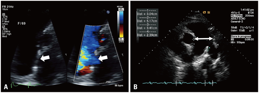
Fig. 1 A: Left: Secundum atrial septal defect in 2-dimensional transthoracic echocardiography (white arrow). Right: Color Doppler flow from left to right atrium (white arrow). B: Dilated main pulmonary artery (white arrow).
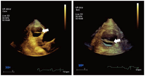
Fig. 2 Left: Bicuspid pulmonic valve (BPV) in 3-dimensional transeso-phageal echocardiography (white arrow). Right: Magnified view of BPV (white arrow).
Reference
-
1. Nair V, Thangaroopan M, Cunningham KS, Mohammed SB, Siu S, Williams WG, Butany J. A bicuspid pulmonary valve associated with tetralogy of fallot. J Card Surg. 2006; 21:185–187.
Article2. Kemaloğlu Öz T, Karadeniz FÖ, Gundlapalli H, Erer B, Sharma RK, Ahmed M, Nanda NC, Yıldırım A, Orhan G, Öz A, Eren M. Coexisting bicuspid aortic and pulmonary valves with normally related great vessels diagnosed by live/real time three-dimensional transesophageal echocardiography. Echocardiography. 2014; 31:218–221.
Article
- Full Text Links
-
- Actions
-
Cited
- CITED
-
- Close
- Share
-
- Similar articles
-
- Clinical and Echocardiographic Features of Pulmonic Valve Endocarditis in patients with Ventricular Septal Defect
- Comparable Outcomes of Bicuspid Aortic Valves for RapidDeployment Aortic Valve Replacement
- A Case of Staphylococcal Tricuspid Valve Endocarditis With Para-Aortic Abscess in a Patient With Bicuspid Aortic Valve
- Mitral Valve Replacement with a Pulmonic Autograft
- Doppler Echocardiographic Assessment of Pre-& Post-Operative Peak Velocity Changes of Four Cardiac Valves in the Left to Right Shunt Lesions
