Enamel renal syndrome with associated amelogenesis imperfecta, nephrolithiasis, and hypocitraturia: A case report
- Affiliations
-
- 1Department of Conservative Dentistry and Endodontics, Manubhai Patel Dental College, Maharaja Krishnakumarsinhji Bhavnagar University, Vadodara, India. aroraankit24@gmail.com
- KMID: 2045026
- DOI: http://doi.org/10.5624/isd.2015.45.3.181
Abstract
- Numerous cases of enamel renal syndrome have been previously reported. Various terms, such as enamel renal syndrome, amelogenesis imperfecta and gingival fibromatosis syndrome, and enamel-renal-gingival syndrome, have been used for patients presenting with the dental phenotype characteristic of this condition, nephrocalcinosis or nephrolithiasis, and gingival findings. This report describes a case of amelogenesis imperfecta of the enamel agenesis variety with nephrolithiasis in a 21-year-old male patient who complained of small teeth. The imaging modalities employed were conventional radiography, cone-beam computed tomography, and renal sonography. Such cases are first encountered by dentists, as other organ or metabolic diseases are generally hidden. Hence, cases of amelogenesis imperfecta should be subjected to advanced diagnostic modalities, incorporating both dental and medical criteria, in order to facilitate comprehensive long-term management.
MeSH Terms
Figure
-

Fig. 1 A. A frontal view shows pigmented gingiva, the absence of enamel with spacing between the teeth, and a semi-lunar appearance of the maxillary central incisors. A right lateral view (B), left lateral view (C), and maxillary and mandibular occlusal views (D and E) show the presence of decayed teeth, molars with a flat occlusal surface, multiple deciduous teeth, and multiple missing permanent teeth. F. An extraoral photograph shows the evidence of wear of anterior teeth.
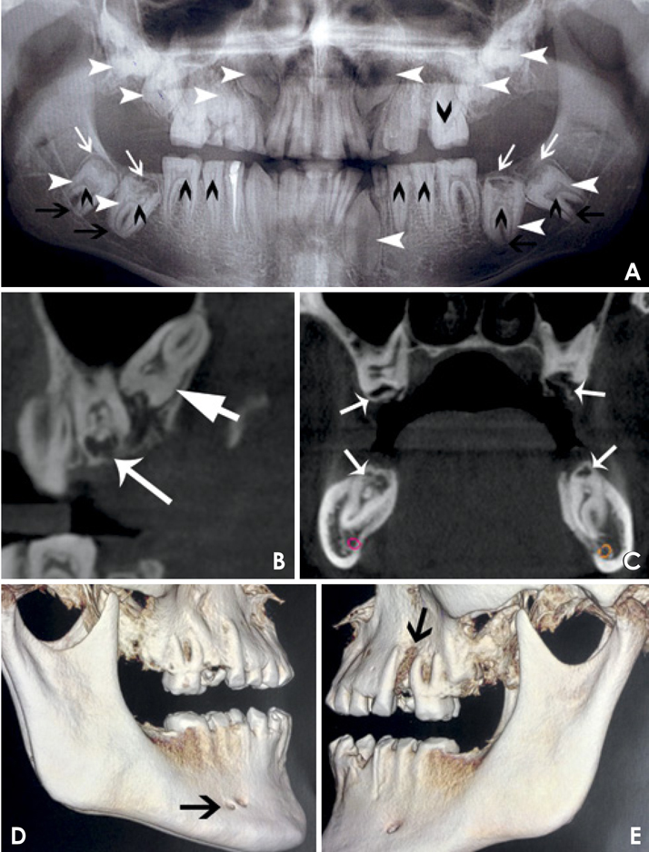
Fig. 2 A. A panoramic radiograph shows pulpal calcifications in most of the teeth (short black arrow), impacted teeth (short white arrows), pericoronal radiolucencies with a sclerotic border (long white arrows), and complete root formation with curvatures (long black arrows). B. A cone-beam CT (CBCT) sagittal image shows uniform radiodensity in the crown (long white arrow) of the dental follicle (long white arrow). C. A CBCT coronal image shows crown resorption (white arrows). D. A CBCT volume-rendered image of the right side of the mandible shows an accessory mental foramen. E. A CBCT volume-rendered image of the left side of the maxilla shows severe bone loss.
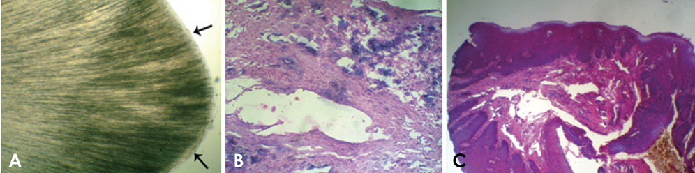
Fig. 3 A. A photomicrograph of a ground section shows the absence of enamel and scalloping of the dentinoenamel junction (black arrows). B. A photomicrograph of the pulpal tissue shows connective tissue with numerous basophilic calcifications (H&E stain, 40×). C. A photomicrograph of the gingival tissue shows parakeratinised epithelium and dense collagen bundles in the lamina propria with fibroblasts and blood vessels (H&E stain, 40×).
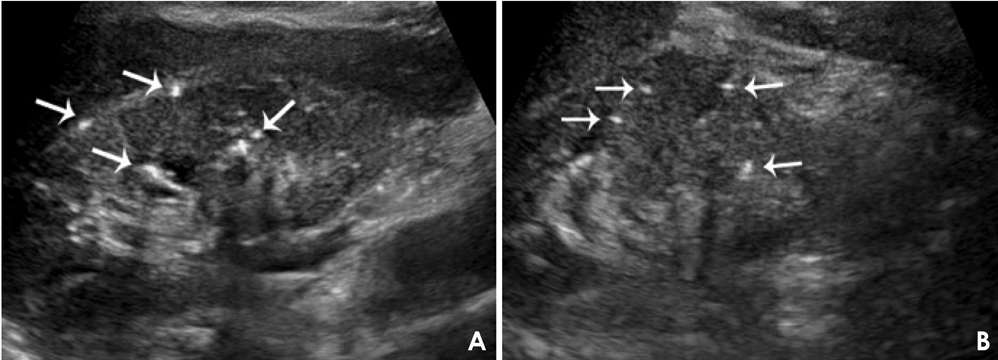
Fig. 4 A and B. Ultrasound images of the left and right kidneys, respectively, show multiple calcified deposits (white arrows).
Reference
-
1. Aldred MJ, Savarirayan R, Crawford PJ. Amelogenesis imperfect: a classification and catalogue for the 21st century. Oral Dis. 2003; 9:19–23.2. Poornima P, Katkade S, Mohamed RN, Mallikarjuna R. Amelogenesis imperfecta with bilateral nephrocalcinosis. BMJ Case Rep. 2013; 2013:bcr2013009370.
Article3. MacGibbon D. Generalized enamel hypoplasia and renal dysfunction. Aust Dent J. 1972; 17:61–63.
Article4. de la Dure-Molla M, Quentric M, Yamaguti PM, Acevedo AC, Mighell AJ, Vikkula M, et al. Pathognomonic oral profile of Enamel Renal Syndrome (ERS) caused by recessive FAM20A Mutations. Orphanet J Rare Dis. 2014; 9:84.
Article5. Kantaputra PN, Kaewgahya M, Khemaleelakul U, Dejkhamron P, Sutthimethakorn S, Thongboonkerd V, et al. Enamel-renalgingival syndrome and FAM20A mutations. Am J Med Genet A. 2014; 164A:1–9.6. Chosack A, Eidelman E, Wisotski I, Cohen T. Amelogenesis imperfecta among Israeli Jews and the description of a new type of local hypoplastic autosomal recessive amelogenesis imperfecta. Oral Surg Oral Med Oral Pathol. 1979; 47:148–156.
Article7. Hashem A, Kelly A, O'Connell B, O'Sullivan M. Impact of moderate and severe hypodontia and amelogenesis imperfecta on quality of life and self-esteem of adult patients. J Dent. 2013; 41:689–694.
Article8. Jaureguiberry G, De la Dure-Molla M, Parry D, Quentric M, Himmerkus N, Koike T, et al. Nephrocalcinosis (enamel renal syndrome) caused by autosomal recessive FAM20A mutations. Nephron Physiol. 2012; 122:1–6.9. Elizabeth J, Lakshmi Priya E, Umadevi KM, Ranganathan K. Amelogenesis imperfecta with renal disease - a report of two cases. J Oral Pathol Med. 2007; 36:625–628.
Article10. Witkop CJ Jr. Amelogenesis imperfecta, dentinogenesis imperfecta and dentin dysplasia revisited: problems in classification. J Oral Pathol. 1988; 17:547–553.
Article11. Crawford PJ, Aldred M, Bloch-Zupan A. Amelogenesis imperfecta. Orphanet J Rare Dis. 2007; 2:17.
Article12. Chamarthi V, Varma BR, Jayanthi M. Amelogenesis imperfecta: a clinician's challenge. J Indian Soc Pedod Prev Dent. 2012; 30:70–73.
Article13. Chaudhary M, Dixit S, Singh A, Kunte S. Amelogenesis imperfecta: report of a case and review of literature. J Oral Maxillofac Pathol. 2009; 13:70–77.
Article14. Hunter L, Addy LD, Knox J, Drage N. Is amelogenesis imperfect an indication for renal examination? Int J Paediatr Dent. 2007; 17:62–65.15. Lubinsky M, Angle C, Marsh PW, Witkop CJ Jr. Syndrome of amelogenesis imperfecta, nephrocalcinosis, impaired renal concentration, and possible abnormality of calcium metabolism. Am J Med Genet. 1985; 20:233–243.
Article16. Del Valle EE, Spivacow FR, Negri AL. Citrate and renal stones. Medicina (B Aires). 2013; 73:363–368.17. Kantaputra PN, Bongkochwilawan C, Kaewgahya M, Ohazama A, Kayserili H, Erdem AP, et al. Enamel-Renal-Gingival syndrome, hypodontia, and a novel FAM20A mutation. Am J Med Genet A. 2014; 164A:2124–2128.18. Martelli-Júnior H, Bonan PR, Dos Santos LA, Santos SM, Cavalcanti MG, Coletta RD. Case reports of a new syndrome associating gingival fibromatosis and dental abnormalities in a consanguineous family. J Periodontol. 2008; 79:1287–1296.
Article19. Udhaya K, Saraladevi KV, Sridhar J. The morphometric analysis of the mental foramen in adult dry human mandibles: a study on the South Indian population. J Clin Diagn Res. 2013; 7:1547–1551.20. Pancer B, Garaicoa-Pazmiño C, Bashutski JD. Accessory mandibular foramen during dental implant placement: case report and review of literature. Implant Dent. 2014; 23:116–124.
- Full Text Links
-
- Actions
-
Cited
- CITED
-
- Close
- Share
-
- Similar articles
-
- Enamel Renal Syndrome: A Case Report of Amelogenesis Imperfecta Associated with Nephrocalcinosis
- Full-mouth rehabilitation in an amelogenesis imperfecta patient with anterior open bite using CAD/CAM system
- Oral rehabilitation of a young adult with amelogenesis imperfect using metal-ceramic restoration: a clinical report
- Multidisciplinary management for amelogenesis imperfecta patient with skeletal CIII malocclusion
- Two Cases of Nephrolithiasis Following Administration of Cyclosporine
