Imaging Sci Dent.
2015 Sep;45(3):175-180. 10.5624/isd.2015.45.3.175.
The reliability of tablet computers in depicting maxillofacial radiographic landmarks
- Affiliations
-
- 1Section of Oral and Maxillofacial Radiology, Department of Oral Health and Diagnostic Sciences, University of Connecticut School of Dental Medicine, Farmington, CT, USA. tadinada@uchc.edu
- 2University of Connecticut School of Dental Medicine, Farmington, CT, USA.
- 3Department of Craniofacial Sciences, University of Connecticut School of Dental Medicine, Farmington, CT, USA.
- 4Section of Operative Dentistry, Department of Reconstructive Sciences, University of Connecticut School of Dental Medicine, Farmington, CT, USA.
- 5Department of Diagnostic Sciences, University of Pittsburgh School of Dentistry, Pittsburgh, PA, USA.
- 6Section of Orthodontics, Department of Craniofacial Sciences, University of Connecticut School of Dental Medicine, Farmington, CT, USA.
- KMID: 2045025
- DOI: http://doi.org/10.5624/isd.2015.45.3.175
Abstract
- PURPOSE
This study was performed to evaluate the reliability of the identification of anatomical landmarks in panoramic and lateral cephalometric radiographs on a standard medical grade picture archiving communication system (PACS) monitor and a tablet computer (iPad 5).
MATERIALS AND METHODS
A total of 1000 radiographs, including 500 panoramic and 500 lateral cephalometric radiographs, were retrieved from the de-identified dataset of the archive of the Section of Oral and Maxillofacial Radiology of the University Of Connecticut School Of Dental Medicine. Major radiographic anatomical landmarks were independently reviewed by two examiners on both displays. The examiners initially reviewed ten panoramic and ten lateral cephalometric radiographs using each imaging system, in order to verify interoperator agreement in landmark identification. The images were scored on a four-point scale reflecting the diagnostic image quality and exposure level of the images.
RESULTS
Statistical analysis showed no significant difference between the two displays regarding the visibility and clarity of the landmarks in either the panoramic or cephalometric radiographs.
CONCLUSION
Tablet computers can reliably show anatomical landmarks in panoramic and lateral cephalometric radiographs.
MeSH Terms
Figure
-
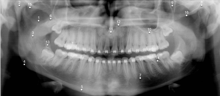
Fig. 1 Anatomical landmarks evaluated on the panoramic radiographs. 1, sigmoid notch; 2, mandibular condyle; 3, mandibular ramus; 4, angle of the mandible; 5, inferior border of the mandible; 6, coronoid process; 7, mental foramen; 8, glenoid fossa; 9, pterygomaxillary fissure; 10, floor of the maxillary sinus; 11, anterior nasal spine; 12, nasal fossa; 13, external oblique ridge; 14, alveolar crest; 15, articular eminence; 16, zygomatic arch.
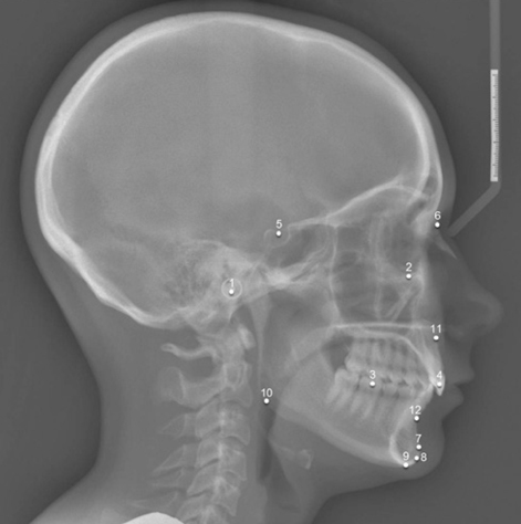
Fig. 2 Anatomical landmarks evaluated on the lateral cephalometric radiographs. 1, porion; 2, orbitale; 3, molars; 4, incisors; 5, sella; 6, nasion; 7, pogonion; 8, gnathion; 9, menton; 10, gonion; 11, point A; 12, point B.
Reference
-
1. Boonn WW, Flanders AE. Informatics in radiology (infoRAD): survey of personal digital assistant use in radiology. Radiographics. 2005; 25:537–541.2. Abboud S, Weiss F, Siegel E, Jeudy J. TB or Not TB: interreader and intrareader variability in screening diagnosis on an iPad versus a traditional display. J Am Coll Radiol. 2013; 10:42–44.
Article3. St Noble V, Thomas R, Gill S, Fascia D. The iPad in portable imaging: radiology on the move. Br J Hosp Med (Lond). 2012; 73:426–427.4. Tahir HJ, Murray IJ, Parry NR, Aslam TM. Optimisation and assessment of three modern touch screen tablet computers for clinical vision testing. PLoS One. 2014; 9:e95074.
Article5. John S, Poh AC, Lim TC, Chan EH, Chong le R. The iPad tablet computer for mobile on-call radiology diagnosis? Auditing discrepancy in CT and MRI reporting. J Digit Imaging. 2012; 25:628–634.
Article6. Johnson PT, Zimmerman SL, Heath D, Eng J, Horton KM, Scott WW, et al. The iPad as a mobile device for CT display and interpretation: diagnostic accuracy for identification of pulmonary embolism. Emerg Radiol. 2012; 19:323–327.
Article7. Shintaku WH, Scarbecz M, Venturin JS. Evaluation of interproximal caries using the iPad 2 and a liquid crystal display monitor. Oral Surg Oral Med Oral Pathol Oral Radiol. 2012; 113:e40–e44.
Article8. White SC, Pharoah MJ. Oral radiology: principles and interpretation. 6th ed. St. Louis: Mosby;2009. p. 200–209.9. Cha JH, Moon WK, Cho N, Lee EH, Park JS, Jang MJ. LCD versus CRT monitors for digital mammography: a comparison of observer performance for the detection of clustered microcalcifications and masses. Acta Radiol. 2009; 50:1104–1108.
Article10. Ikeda R, Katsuragawa S, Shimonobou T, Hiai Y, Hashida M, Awai K, et al. Comparison of LCD and CRT monitors for detection of pulmonary nodules and interstitial lung diseases on digital chest radiographs by using receiver operating characteristic analysis. Nihon Hoshasen Gijutsu Gakkai Zasshi. 2006; 62:734–741.
Article11. Kallio-Pulkkinen S, Haapea M, Liukkonen E, Huumonen S, Tervonen O, Nieminen MT. Comparison of consumer grade, tablet and 6MP-displays: observer performance in detection of anatomical and pathological structures in panoramic radiographs. Oral Surg Oral Med Oral Pathol Oral Radiol. 2014; 118:135–141.
Article12. Hellén-Halme K, Nilsson M, Petersson A. Effect of monitors on approximal caries detection in digital radiographs - standard versus precalibrated DICOM part 14 displays: an in vitro study. Oral Surg Oral Med Oral Pathol Oral Radiol Endod. 2009; 107:716–720.13. Hellén-Halme K, Petersson A, Warfvinge G, Nilsson M. Effect of ambient light and monitor brightness and contrast settings on the detection of approximal caries in digital radiographs: an in vitro study. Dentomaxillofac Radiol. 2008; 37:380–384.14. Hellén-Halme K, Lith A. Carious lesions: diagnostic accuracy using pre-calibrated monitor in various ambient light levels: an in vitro study. Dentomaxillofac Radiol. 2013; 42:20130071.15. Yoshimura K, Shimamoto K, Ikeda M, Ichikawa K, Naganawa S. A comparative contrast perception phantom image of brain CT study between high-grade and low-grade liquid crystal displays (LCDs) in electronic medical charts. Phys Med. 2011; 27:109–116.
Article16. Baheti MJ, Toshniwal N. Orthodontic apps at fingertips. Prog Orthod. 2014; 15:36.
Article
- Full Text Links
-
- Actions
-
Cited
- CITED
-
- Close
- Share
-
- Similar articles
-
- A comparative study of computed radiographic cephalometry and conventional cephalometry in reliability of head film measurements (landmarks identification)
- Radiographic examination for successful dental implant
- Reproducibility of Lateral Cephalometric Landmarks According to Radiographic Image Enhancement
- Evaluation of the accuracy and reliability of 3-dimensional computerized tomography for measurement of maxillofacial region
- Comparison of the observer reliability of cranial anatomic landmarks based on cephalometric radiograph and three-dimensional computed tomography scans
