J Korean Assoc Oral Maxillofac Surg.
2010 Aug;36(4):262-269. 10.5125/jkaoms.2010.36.4.262.
Comparison of the observer reliability of cranial anatomic landmarks based on cephalometric radiograph and three-dimensional computed tomography scans
- Affiliations
-
- 1Department of Oral and Maxillofacial Surgery, Sun Dental Hospital, Daejeon, Korea.
- 2Department of Oral and Maxillofacial Surgery, School of Dentistry, Kyungpook National University, Daegu, Korea. shalee@knu.ac.kr
- KMID: 1457777
- DOI: http://doi.org/10.5125/jkaoms.2010.36.4.262
Abstract
- INTRODUCTION
Accurate diagnosis and treatment planning are very important for orthognathic surgery. A small error in diagnosis can cause postoperative functional and esthetic problems. Pre-existing 2-dimensional (D) chephalogram analysis has a high likelihood of error due to its intrinsic and extrinsic problems. A cephalogram can also be inaccurate due to the limited anatomic points, superimposition of the image, and the considerable time and effort required. Recently, an improvement in technology and popularization of computed tomography (CT) provides patients with 3-D computer based cephalometric analysis, which complements traditional analysis in many ways. However, the results are affected by the experience and the subject of the investigator.
MATERIALS AND METHODS
The effects of the sources human error in 2-D cephalogram analysis and 3-D computerized tomography cephalometric analysis were compared using Simplant CMF program. From 2008 Jan to 2009 June, patients who had undergone CT, cephalo AP, lat were investigated.
RESULTS
1. In the 3 D and 2 D images, 10 out of 93 variables (10.4%) and 11 out 44 variables (25%), respectively, showed a significant difference. 2. Landmarks that showed a significant difference in the 2 D image were the points frequently superimposed anatomically. 3. Go Po Orb landmarks, which showed a significant difference in the 3 D images, were found to be the artificial points for analysis in the 2 D image, and in the current definition, these points cannot be used for reproducibility in the 3 D image.
CONCLUSION
Generally, 3-D CT images provide more precise identification of the traditional cephalometric landmark. Greater variability of certain landmarks in the mediolateral direction is probably related to the inadequate definition of the landmarks in the third dimension.
Keyword
MeSH Terms
Figure
-

Fig. 1. Two-dimensional analysis of V-Ceph (version 4.0): cephalometric lateral view.
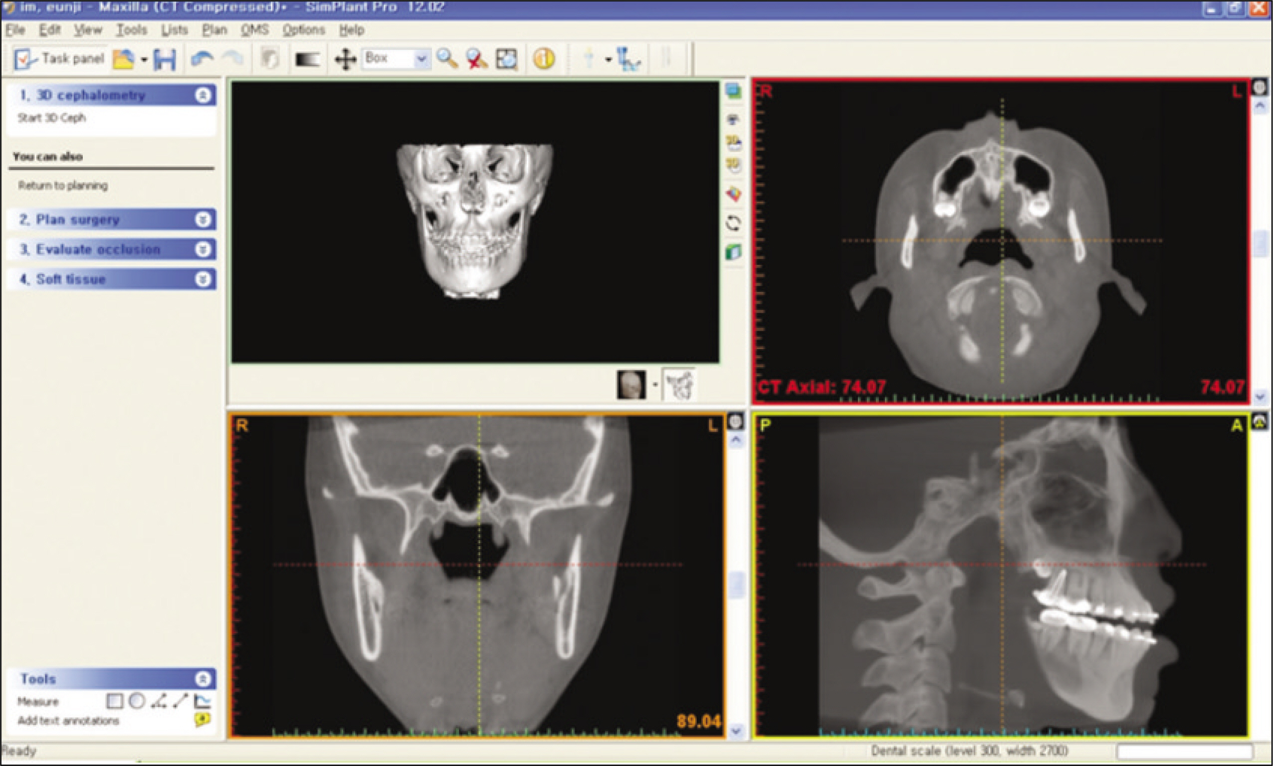
Fig. 2. Three-dimensional analysis of SimPlant Pro (version 12.02): 3-dimentional image and axial, coronal, sagittal view.

Fig. 3. Coronal and sagittal view: sagittal plane makes using 3 point (nasion, ANS, dens of axis) coronal plane makes using porion. (ANS: anterior nasal spine)

Fig. 4. Skeletal landmark distance: measure from 3 standard plane to point.
Cited by 1 articles
-
Comparison of landmark positions between Cone-Beam Computed Tomogram (CBCT) and Adjusted 2D lateral cephalogram
Soo-Jung Son, Youn-Sic Chun, Minji Kim
J Korean Acad Prosthodont. 2014;52(3):222-232. doi: 10.4047/jkap.2014.52.3.222.
Reference
-
References
1. Grayson BH, McCarthy JG, Bookstein F. Analysis of craniofacial asymmetry by multiplane cephalometry. Am J Orthod. 1983; 84:217–24.
Article2. Baumrind S, Moffitt FH, Curry S. Three-dimensional x-ray stereometry from paired coplanar images: a progress report. Am J Orthod. 1983; 84:292–312.
Article3. Baumrind S, Moffitt FH, Curry S. The geometry of three-dimensional measurement from paired coplanar x-ray images. Am J Orthod. 1983; 84:313–22.
Article4. Moss JP, Linney AD, Grindrod SR, Arridge SR, Clifton JS. Three-dimensional visualization of the face and skull using computerized tomography and laser scanning techniques. Eur J Orthord. 1987; 9:247–53.
Article5. Grayson B, Cutting C, Bookstein FL, Kim H, McCarthy JG. The three-dimensional cephalogram: theory, technique, and clinical application. Am J Orthod Dentofacial Orthop. 1988; 94:327–37.6. Bookstein FL, Grayson B, Cutting CB, Kim HC, McCarthy JG. Landmarks in three dimension: recontruction from cephalograms versus direct observation. Am J Orthord Dentofacial Orthop. 1991; 100:133–40.7. Ferrario VF, Sforza C, Poggio CE, Tartaglia G. Distance from symmetry: a three-dimensional evaluation of facial asymmetry. J Oral Maxillofac Surg. 1994; 52:1126–32.
Article8. Ferrario VF, Sforza C, Puleo A, Poggio CE, Schmitz JH. Three-dimensional facial morphometry and conventional cephalometrics: a correlation study. Int J Adult Orthodon Orthognath Surg. 1996; 11:329–38.9. Kusnoto B, Evans CA, BeGole EA, de Rijk W. Assessment of 3-dimensional computer-generated cephalometric measurement. Am J Orthod Dentofacial Orthop. 1999; 116:390–9.10. Cavalcanti MG. Vannier MW. Quantitative analysis of spiral computed tomography for craniofacial clinical applications. Dentomaxillofac Radiol. 1998; 27:344–50.11. Nawaratne S, Fabiny R, Brien JE, Zalcberg J, Cosolo W, Whan A, et al. Accuracy of volume measurement using helical CT. J Comput Assist Tomogr. 1997; 21:481–6.
Article12. Xia J, lp HH, Samman N, Wang D, Kot CS, Yeung RW, et al. Computer-assisted three-dimensional surgical planning and stimulation 3D virtual osteotomy. Int J Oral Maxillofac Surg. 2000; 29:11–7.13. Katsumata A, Fujishita M, Maeda M, Ariji Y, Ariji E, Langlais RP. 3D-CT evaluation of facial asymmetry. Oral Surg Oral Med Oral Pathol Oral Radiol Endod. 2005; 99:212–20.
Article14. Kawamata A, Ariji Y, Langlais RP. Three-dimensional computed tomography imaging in dentistry. Dent Clin North Am. 2000; 44:395–440.15. Kawamata A, Ariji Y, Langlais RP. Three-dimensional imaging for orthognathic surgery and orthodontic treatment. Oral Maxillofac Surg Clin North Am. 2001; 13:713–25.
Article16. Matteson SR, Bechtold W, Phillips C, Staab EV. A method for three dimensional image reformation for quantitative cephalometric analysis. J Oral Maxillofac Surg. 1989; 47:1053–61.17. Hildebolt CF, Vannier MW, Knapp RH. Validation study of skull three-dimensional computerized tomography measurements. Am J Phys Anthropol. 1990; 82:283–94.
Article18. Cavalcanti MG, Haller JW, Vannier MW. Three-dimensional computed tomography landmark measurement in craniofacial surgical planning experimental validation in vitro. J Oral Maxillofac Surg. 1999; 57:690–4.
Article19. Song JC, Jang HJ, Lee SH. Evaluation of the accuracy and reliability of 3-dimensional computerized tomography for measurement of maxillofacial region. J Korean Assoc Maxillofac Plast Reconstr Surg. 2000; 22:500–12.20. Christiansen EL, Thompson JR, Kopp S. Intra-and interobserver variability and accuracy in the determination of linear and angular measurements in computed tomography. An in vitro and in situ study of human mandibles. Acta Odontol Scand. 1986; 44:221–9.21. Richtsmeier JT, Paik CH, Elfert PC, Cole TM, Dahlman HR. Precision, repeatabillity and validation of the localization of cranial landmarks using computed tomography scans. Cleft Palate Craniofac J. 1995; 32:217–27.22. Kragskov J, Bosch C, Gyldensted C, Sindet-Pedersen S. Comparison of the reliability of craniofacial anatomic landmarks based on cephalometric radiographs and three-dimensional CT scans. Cleft Palate Craniofac J. 1997; 34:111–6.
Article23. Papadopoulos MA, Jannowitz C, Boettcher P, Henke J, Stolla R, Zeilhofer HF, et al. Three-dimensional fetal cephalometry: an evaluation of the reliability of cephalometric measurements based on three-dimensional CT reconstructions and on dry skulls of sheep fetuses. J Craniomaxillofac Surg. 2005; 33:229–37.
Article24. Midtgard J, Bjork G, Linder-Aronson S. Reproducibility of cephalometric landmarks and errors of measurements of cephalometric cranial distances. Angle Orthod. 1974; 44:56–61.25. Ha ¨ gg U, Cooke MS, Chan TC, Tng TT, Lau PY. The reproducibility of cephalometric landmarks: an experimental study on skulls. Aust Orthod J. 1998; 15:177–85.
- Full Text Links
-
- Actions
-
Cited
- CITED
-
- Close
- Share
-
- Similar articles
-
- Evaluation of the accuracy and reliability of 3-dimensional computerized tomography for measurement of maxillofacial region
- Inter- and Intra-observer Variability of a Cervical OPLL Classification Using Reconstructed CT Images
- Reproducibility of Lateral Cephalometric Landmarks According to Radiographic Image Enhancement
- A comparative study of computed radiographic cephalometry and conventional cephalometry in reliability of head film measurements (landmarks identification)
- Comparative study of glenoid version and inclination using two-dimensional images from computed tomography and three-dimensional reconstructed bone models
