A comparative study of metal artifacts from common metal orthodontic brackets in magnetic resonance imaging
- Affiliations
-
- 1Department of Maxillofacial Radiology, Faculty of Dentistry, Guilan University of Medical Sciences, Rasht, Iran. zahradalili@yahoo.com
- 2Department of Orthodontics, Faculty of Dentistry, Guilan University of Medical Sciences, Rasht, Iran.
- 3Department of Radiology, Poursina Hospital, Guilan University of Medical Sciences, Rasht, Iran.
- 4Faculty of Dentistry, Guilan University of Medical Sciences, Rasht, Iran.
- 5Department of Biostatistics, Guilan University of Medical Sciences, Rasht, Iran.
- KMID: 2045023
- DOI: http://doi.org/10.5624/isd.2015.45.3.159
Abstract
- PURPOSE
This study was performed to compare the metal artifacts from common metal orthodontic brackets in magnetic resonance imaging.
MATERIALS AND METHODS
A dry mandible with 12 intact premolars was prepared, and was scanned ten times with various types of brackets: American, 3M, Dentaurum, and Masel orthodontic brackets were used, together with either stainless steel (SS) or nickel titanium (NiTi) wires. Subsequently, three different sequences of coronal and axial images were obtained: spin-echo T1-weighted images, fast spin-echo T2-weighted images, and fluid-attenuated inversion recovery images. In each sequence, the two sequential axial and coronal images with the largest signal-void area were selected. The largest diameters of the signal voids in the direction of the X-, Y-, and Z-axes were then measured twice. Finally, the mean linear values associated with different orthodontic brackets were analyzed using one-way analysis of variation, and the results were compared using the independent t-test to assess whether the use of SS or NiTi wires had a significant effect on the images.
RESULTS
Statistically significant differences were only observed along the Z-axis among the four different brands of orthodontic brackets with SS wires. A statistically significant difference was observed along all axes among the brackets with NiTi wires. A statistically significant difference was found only along the Z-axis between nickel-free and nickel-containing brackets.
CONCLUSION
With respect to all axes, the 3M bracket was associated with smaller signal-void areas. Overall, the 3M and Dentaurum brackets with NiTi wires induced smaller artifacts along all axes than those with SS wires.
MeSH Terms
Figure
-
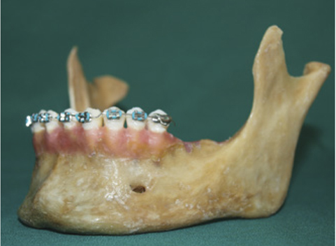
Fig. 1 The brackets are bonded with composite onto the teeth for performing magnetic resonance imaging.
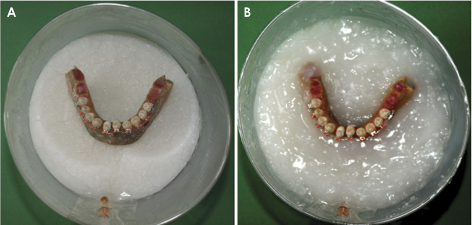
Fig. 2 A and B. The mandible is fixed in the starch mixture.
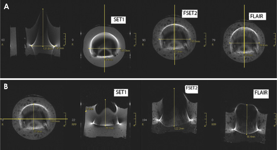
Fig. 3 Axial (A) and coronal (B) images reveal various measurements of signal-void areas along the X-, Y-, and Z-axes detected in the different imaging sequences for American-brand brackets with NiTi wire.
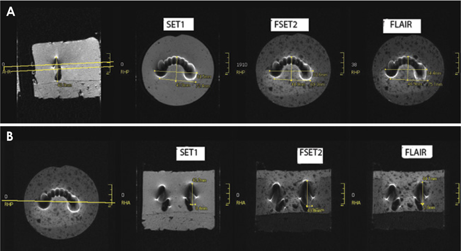
Fig. 4 Axial (A) and coronal (B) views reveal the various measurements of the signal-void areas along the X-, Y-, and Z-axes in the different imaging sequences for Dentaurum-brand brackets with nickel titanium wires.
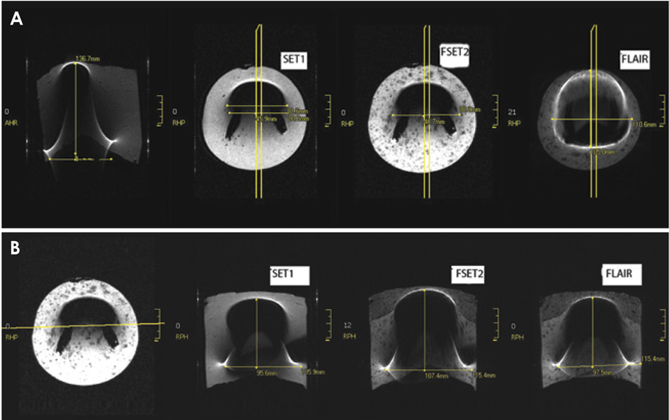
Fig. 5 Axial (A) and coronal (B) views show the various measurements of the signal-void areas found along the X-, Y-, and Z-axes in the different imaging sequences for Masel-brand brackets with stainless steel wires.
Reference
-
1. Shafiei F, Honda E, Takahashi H, Sasaki T. Artifacts from dental casting alloys in magnetic resonance imaging. J Dent Res. 2003; 82:602–606.2. Idiyatullin D, Corum C, Moeller S, Prasad HS, Garwood M, Nixdorf DR. Dental magnetic resonance imaging: making the invisible visible. J Endod. 2011; 37:745–752.
Article3. Behr M, Fellner C, Bayreuther G, Leibrock A, Held P, Fellner F, et al. MR-imaging of the TMJ: artefacts caused by dental alloys. Eur J Prosthodont Restor Dent. 1996; 4:111–115.4. Hargreaves BA, Worters PW, Pauly KB, Pauly JM, Koch KM, Gold GE. Metal-induced artifacts in MRI. AJR Am J Roentgenol. 2011; 197:547–555.
Article5. Bennett LH, Wang PS, Donahue MJ. Artifacts in magnetic resonance imaging from metals. J Appl Phys. 1996; 79:4712–4714.
Article6. Bui FM, Bott K, Mintchev MP. A quantitative study of the pixel-shifting, blurring and nonlinear distortions in MRI images caused by the presence of metal implants. J Med Eng Technol. 2000; 24:20–27.7. Sadowsky PL, Bernreuter W, Lakshminarayanan AV, Kenney P. Orthodontic appliances and magnetic resonance imaging of the brain and temporomandibular joint. Angle Orthod. 1988; 58:9–20.8. Pusey E, Lufkin RB, Brown RK, Solomon MA, Stark DD, Tarr RW, et al. Magnetic resonance imaging artifacts: mechanism and clinical significance. Radiographics. 1986; 6:891–911.
Article9. Hopper TA, Vasilic B, Pope JM, Jones CE, Epstein CL, Song HK, et al. Experimental and computational analyses of the effects of slice distortion from a metallic sphere in an MRI phantom. Magn Reson Imaging. 2006; 24:1077–1085.
Article10. Klinke T, Daboul A, Maron J, Gredes T, Puls R, Jaghsi A, et al. Artifacts in magnetic resonance imaging and computed tomography caused by dental material. PLoS One. 2012; 7:e31766.11. Hajnal JV, Bryant DJ, Kasuboski L, Pattany PM, De Coene B, Lewis PD, et al. Use of fluid attenuated inversion recovery (FLAIR) pulse sequences in MRI of the brain. J Comput Assist Tomogr. 1992; 16:841–844.
Article12. Costa AL, Appenzeller S, Yasuda CL, Pereira FR, Zanardi VA, Cendes F. Artifacts in brain magnetic resonance imaging due to metallic dental objects. Med Oral Patol Oral Cir Bucal. 2009; 14:E278–E282.13. Hasegawa M, Miyata K, Abe Y, Ishigami T. Radiofrequency heating of metallic dental devices during 3.0 T MRI. Dentomaxillofac Radiol. 2013; 42:20120234.14. Farahani K, Sinha U, Sinha S, Chiu LC, Lufkin RB. Effect of field strength on susceptibility artifacts in magnetic resonance imaging. Comput Med Imaging Graph. 1990; 14:409–413.
Article15. Harris TM, Faridrad MR, Dickson JA. The benefits of aesthetic orthodontic brackets in patients requiring multiple MRI scanning. J Orthod. 2006; 33:90–94.
Article16. Shellock FG, Kanal E. Aneurysm clips: evaluation of MR imaging artifacts at 1.5 T. Radiology. 1998; 209:563–566.
Article17. Okano Y, Yamashiro M, Kaneda T, Kasai K. Magnetic resonance imaging diagnosis of the temporomandibular joint in patients with orthodontic appliances. Oral Surg Oral Med Oral Pathol Oral Radiol Endod. 2003; 95:255–263.
Article18. Morikuni H, Koseki T, Yotsui Y, Matsumoto N. Metallic artifacts on magnetic resonance images caused by multibracket appliances. J Osaka Dent Univ. 2010; 44:13–21.19. Patel A, Bhavra GS, O'Neill JR. MRI scanning and orthodontics. J Orthod. 2006; 33:246–249.
Article20. Kemper J, Priest AN, Schulze D, Kahl-Nieke B, Adam G, Klocke A. Orthodontic springs and auxilliary appliance: assessment of magnetic field interactions associated with 1.5 T and 3 T magnetic resonance systems. Eur Radiol. 2007; 17:533–540.
- Full Text Links
-
- Actions
-
Cited
- CITED
-
- Close
- Share
-
- Similar articles
-
- A study on frictional resistance force of orthodontic resin bracket
- Shear bond strength of metal brackets bonded with light-cured adhesive: an in vitro comparative study
- Orthodontic appliances and MR image artefacts: An exploratory in vitro and in vivo study using 1.5-T and 3-T scanners
- In vitro physical, chemical, and biological evaluation of commercially available metal orthodontic brackets
- A scanning electron microscopic study on the corrosion resistance of chemically and thermally recycled metal brackets
