In vitro physical, chemical, and biological evaluation of commercially available metal orthodontic brackets
- Affiliations
-
- 1GIO Dental Clinic, Gimpo, Korea.
- 2Department of Orthodontics, College of Dentistry, Institute of Craniofacial Deformity, Yonsei University, Seoul, Korea. hwang@yuhs.ac
- KMID: 1976682
- DOI: http://doi.org/10.4041/kjod.2012.42.6.297
Abstract
OBJECTIVE
This in vitro study was undertaken to evaluate the physical, chemical, and biological properties of commercially available metal orthodontic brackets in South Korea, because national standards for these products are lacking.
METHODS
Four bracket brands were tested for dimensional accuracy, (manufacturing errors in angulation and torque), cytotoxicity, composition, elution, and corrosion: Archist (Daeseung Medical), Victory (3M Unitek), Kosaka (Tomy), and Confidence (Shinye Odontology Materials).
RESULTS
The tested rackets showed no significant differences in manufacturing errors in angulation, but Confidence brackets showed a significant difference in manufacturing errors in torque. None of the brackets were cytotoxic to mouse fibroblasts. The metal ion components did not show a regular increasing or decreasing trend of elution over time, but the volume of the total eluted metal ions increased: Archist brackets had the maximal Cr elution and Confidence brackets appeared to have the largest volume of total eluted metal ions because of excessive Ni elution. Confidence brackets showed the lowest corrosion resistance during potentiodynamic polarization.
CONCLUSIONS
The results of this study could potentially be applied in establishing national standards for metal orthodontic brackets and in evaluating commercially available products.
Keyword
Figure
-
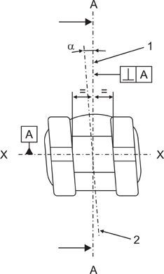
Figure 1 Measurement of angulation (α). X, Mesiodistal long axis of the slot; 1, line perpendicular to X; 2, central occlusogingival axis.
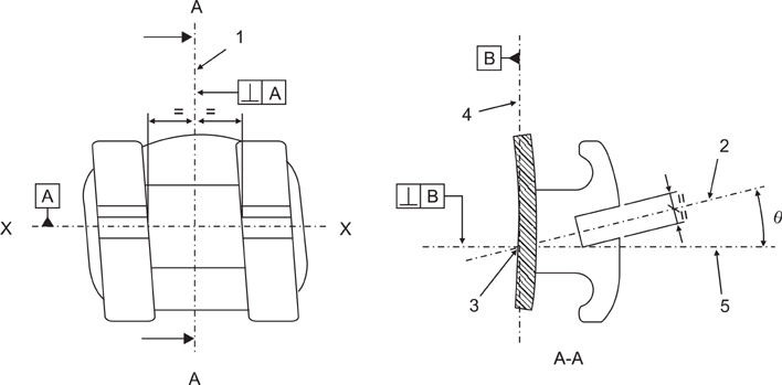
Figure 2 Measurement of torque. X, Mesiodistal long axis of the slot; 1, plane perpendicular to X in the centre of the slot; 2, line bisecting the slot in the occlusogingival direction; 3, intersection of line 2 and the tooth side surface of the base in plane 1; 4, tangent to the base at 3; 5, line perpendicular to line 4 at 3; θ, angle of torque.

Figure 3 Preparation of cells and specimens for the cytotoxicity test.
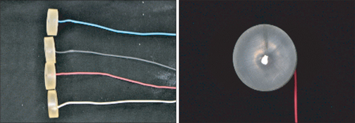
Figure 4 Samples and their respective bases for the corrosion test.
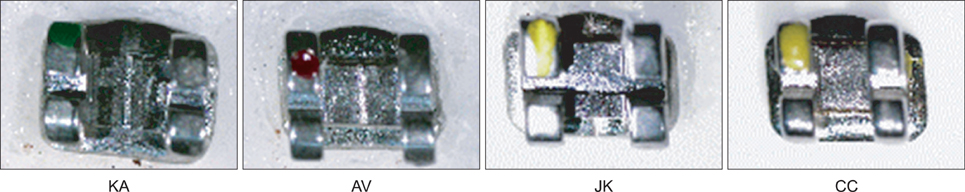
Figure 5 Frontal view of the tested brackets. KA, Korea-Archist; AV, America-Victory; JK, Japan-Kosaka; CC, China-Confidence.
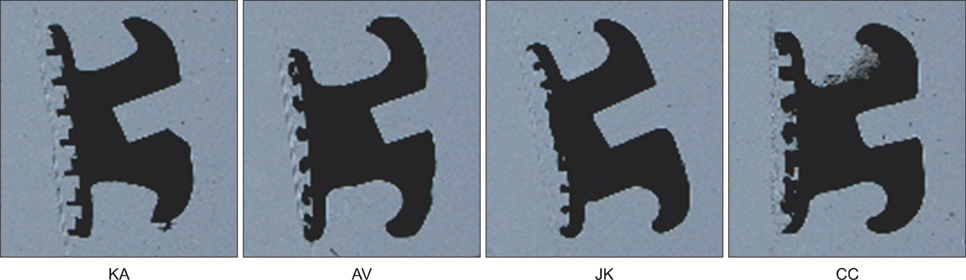
Figure 6 Cross-sectional view of the tested brackets. KA, Korea-Archist; AV, America-Victory; JK, Japan-Kosaka; CC, China-Confidence.
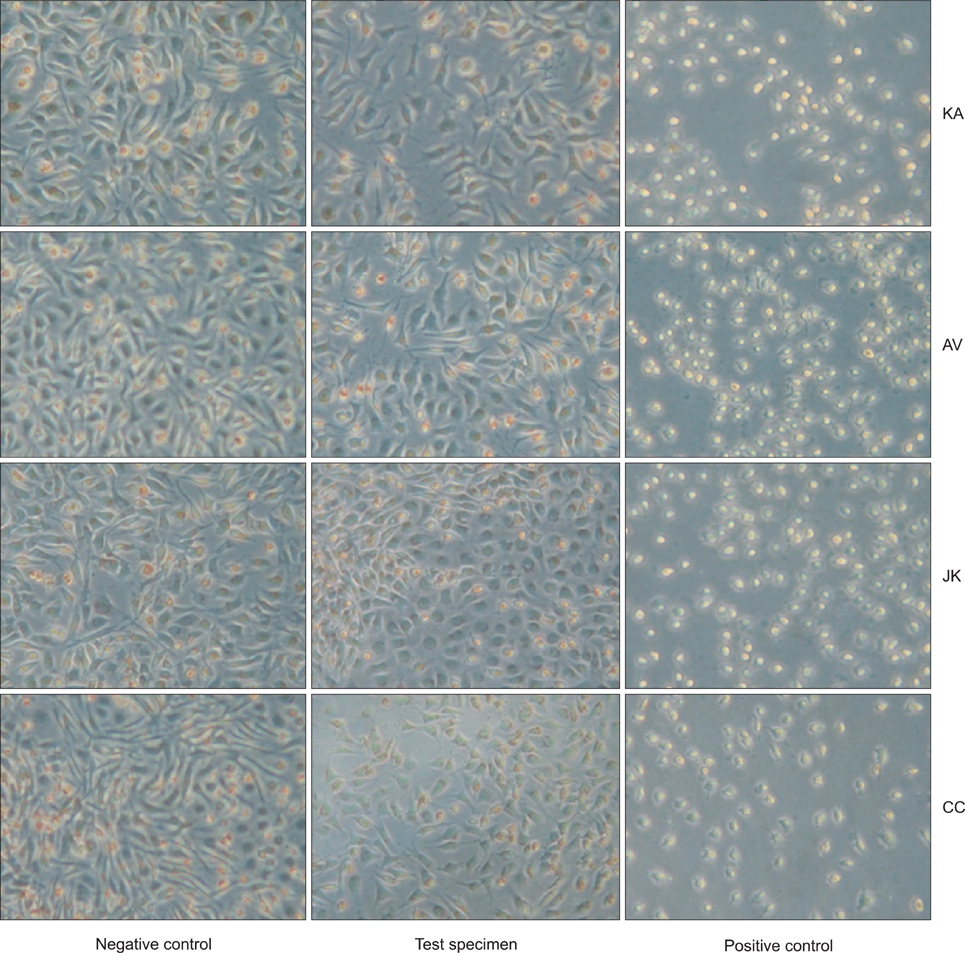
Figure 7 Appearance of cells around the specimens in the cytotoxicity test. KA, Korea-Archist; AV, America-Victory; JK, Japan-Kosaka; CC, China-Confidence.
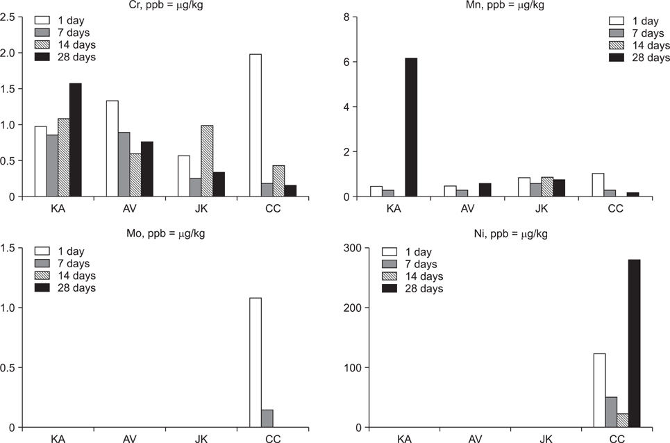
Figure 8 Volume of metal ions released according to elapsed time. KA, Korea-Archist; AV, America-Victory; JK, Japan-Kosaka; CC, China-Confidence.
Reference
-
1. Creekmore TD, Kunik RL. Straight wire: the next generation. Am J Orthod Dentofacial Orthop. 1993. 104:8–20.
Article2. Ødegaard J, Segner D. Shear bond strength of metal brackets compared with a new ceramic bracket. Am J Orthod Dentofacial Orthop. 1988. 94:201–206.
Article3. Feldner JC, Sarkar NK, Sheridan JJ, Lancaster DM. In vitro torque-deformation characteristics of orthodontic polycarbonate brackets. Am J Orthod Dentofacial Orthop. 1994. 106:265–272.
Article4. Cacciafesta V, Sfondrini MF, Scribante A, Klersy C, Auricchio F. Evaluation of friction of conventional and metal-insert ceramic brackets in various bracket-archwire combinations. Am J Orthod Dentofacial Orthop. 2003. 124:403–409.
Article5. Kapur Wadhwa R, Kwon HK, Sciote JJ, Close JM. Frictional resistance in ceramic and metal brackets. J Clin Orthod. 2004. 38:35–38.6. Kim JH, Kwon OW, Choi YY. Discoloration and corrosion resistance of Tin-coated orthodontic appliances produced by Ion-plating method. Korean J Orthod. 1993. 23:327–340.7. Maijer R, Smith DC. Biodegradation of the orthodontic bracket system. Am J Orthod Dentofacial Orthop. 1986. 90:195–198.
Article8. Toms AP. The corrosion of orthodontic wire. Eur J Orthod. 1988. 10:87–97.
Article9. House K, Sernetz F, Dymock D, Sandy JR, Ireland AJ. Corrosion of orthodontic appliances-should we care? Am J Orthod Dentofacial Orthop. 2008. 133:584–592.
Article10. Park HY, Shearer TR. In vitro release of nickel and chromium from simulated orthodontic appliances. Am J Orthod. 1983. 84:156–159.
Article11. Lim YK, Yang WS. An experimental study on the cytotoxicity of orthodontic wires. Korean J Orthod. 1996. 26:591–599.12. Lee GH, Cho JH, Lee KH, Hwang HS. Evaluation of cytotoxicity of electroplated stainless steel orthodontic wire. Korean J Orthod. 2005. 35:127–136.13. Ceen RF, Gwinnett AJ. Indelible iatrogenic staining of enamel following debonding: a case report. J Clin Orthod. 1980. 14:713–715.14. Maijer R, Smith DC. Corrosion of orthodontic bracket bases. Am J Orthod. 1982. 81:43–48.
Article15. Kim MS, Sung JH, Kwon OW. A study on metal release of tin ion-plated stainless steel orthodontic appliances. Korean J Orthod. 1995. 25:43–54.16. International Organization for Standardization. Dentistry: brackets and tubes for use in orthodontics. ISO 27020. 2010.17. International Organization for Standardization. Biological evaluation of medical devices. Part 5: Test for in vitro cytotoxicity. ISO 10993-5. 2009.18. International Organization for Standardization. Dental metallic materials: corrosion test methods. ISO 10271. 2011.19. Oh KT, Choo SU, Kim KM, Kim KN. A stainless steel bracket for orthodontic application. Eur J Orthod. 2005. 27:237–244.
Article20. Kao CT, Ding SJ, Min Y, Hsu TC, Chou MY, Huang TH. The cytotoxicity of orthodontic metal bracket immersion media. Eur J Orthod. 2007. 29:198–203.
Article21. Yoo DH, Kook YA, Kim SC. An experimental study on the cytotoxicity of various orthodontic bands. Korean J Orthod. 1994. 24:419–432.22. Lim YK, Yang WS. An experimental study on the cytotoxicity of recycled brackets. Korean J Orthod. 1993. 23:147–163.23. Oh KT, Kim KN. Ion release and cytotoxicity of stainless steel wires. Eur J Orthod. 2005. 27:533–540.
Article24. Freitas MP, Oshima HM, Menezes LM, Machado DC, Viezzer C. Cytotoxicity of silver solder employed in orthodontics. Angle Orthod. 2009. 79:939–944.
Article25. Eliades T, Athanasiou AE. In vivo aging of orthodontic alloys: implications for corrosion potential, nickel release, and biocompatibility. Angle Orthod. 2002. 72:222–237.26. Staerkjaer L, Menné T. Nickel allergy and orthodontic treatment. Eur J Orthod. 1990. 12:284–289.
Article27. Sfondrini MF, Cacciafesta V, Maffia E, Scribante A, Alberti G, Biesuz R, et al. Nickel release from new conventional stainless steel, recycled, and nickel-free orthodontic brackets: an in vitro study. Am J Orthod Dentofacial Orthop. 2010. 137:809–815.
Article28. Choi CM, Rhee BT. The effects of heat treatment on mechanical properties and metal release from heat-treated orthodontic archwires. Korean J Orthod. 1990. 20:381–390.29. Grimsdottir MR, Gjerdet NR, Hensten-Pettersen A. Composition and in vitro corrosion of orthodontic appliances. Am J Orthod Dentofacial Orthop. 1992. 101:525–532.
Article30. National Research Council. Dietary reference intakes: the essential guide to nutrient requirements. 2006. Washington, DC: The National Academies Press.
- Full Text Links
-
- Actions
-
Cited
- CITED
-
- Close
- Share
-
- Similar articles
-
- Shear bond strength of metal brackets bonded with light-cured adhesive: an in vitro comparative study
- A study on frictional resistance force of orthodontic resin bracket
- Friction of calcium phosphate brackets to stainless steel wire
- In vitro shear bond strength of ceramic brackets
- The effects of fluoride releasing orthodontic sealant on the shear bond strength of light-and chemical-cured orthodontic resins
