J Korean Foot Ankle Soc.
2015 Sep;19(3):102-106. 10.14193/jkfas.2015.19.3.102.
Comparison of the Results between Cadaveric and Radiological Measurements of Calcaneus
- Affiliations
-
- 1Department of Orthopedic Surgery, Busan Paik Hospital, Inje University College of Medicine, Busan, Korea. ortho1@hanmail.net
- 2Department of Orthopaedic Surgery, Hyosung City Hospital, Busan, Korea.
- KMID: 2045013
- DOI: http://doi.org/10.14193/jkfas.2015.19.3.102
Abstract
- PURPOSE
We wanted to compare the results between cadaveric and radiological measurements of calcaneus.
MATERIALS AND METHODS
Sixty three calcaneus of 33 cadavers donated between December 2012 and December 2014 were actually measured. Computed tomography (CT) images of 244 calcaneus in 122 patients of the same age group with cadavers were radiologically measured. Maximum length, maximum width, maximum height, Bohler angle, Gissane angle, Fowler-Phillip angle, and tala-articular angle were measured.
RESULTS
In cadaveric measurement, the mean maximal height, length, and width were 41.8+/-3.3 mm, 73.3+/-3.4 mm, and 40.7+/-2.2 mm, respectively. In radiological measurement, the mean maximal height, length, and width were 38.5+/-4.3 mm, 74.0+/-5.7 mm, and 44.7+/-1.4 mm, respectively. In cadaveric measurement, the mean Bohler angle, Gissane angle, Fowler-Phillip angle, and tala-articular angle were 32.1degrees+/-6.2degrees, 110.8degrees+/-8.1degrees, 55.8degrees+/-6.8degrees, and 59.7degrees+/-4.6degrees, respectively. In radiological measurement the mean Bohler angle, Gissane angle, Fowler-Phillip angle, and tala-articular angle were 32.6degrees+/-3.8degrees, 113.7degrees+/-5.7degrees, 62.2degrees+/-3.9degrees, and 61.6degrees+/-6.3degrees, respectively. The mean maximal height was significantly higher in the cadaveric measurement group (p<0.001) and the mean maximal length and width were significantly higher in the radiologic measurement group (p<0.001, p<0.001). The mean Gissane angle, Fowler-Philip angle, and Bohler angle were significantly higher in the CT group (p=0.001, p<0.001, p=0.016, respectively). There was no significant difference in the mean tala-articular angle (p=0.352).
CONCLUSION
Significant differences in length parameters were observed between the cadaveric measurement group and the radiologic measurement group. However, no significant differences in angular measurements were observed between the two groups. The authors carefully conclude that radiological measurement values may be different from actual values in the calcaneus.
Keyword
MeSH Terms
Figure
-
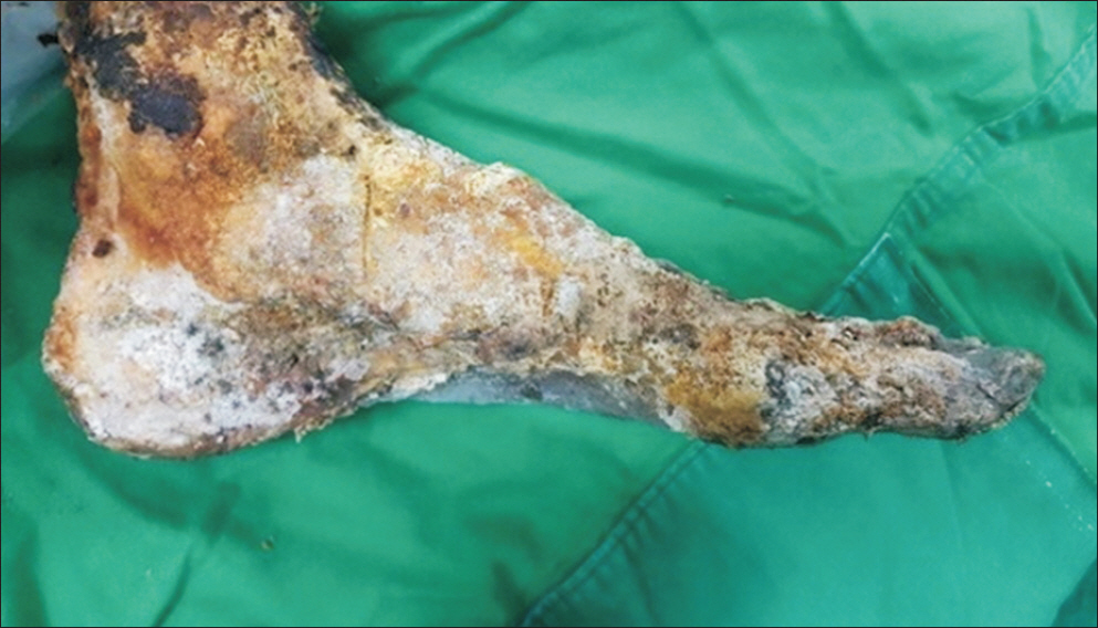
Figure 1. This photograph shows an example of severely decayed cadaver.
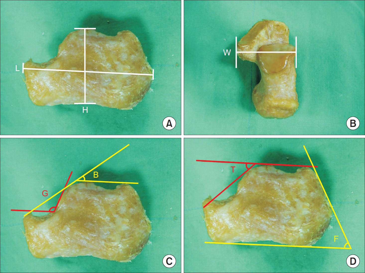
Figure 2. Actual measurement of calcaneus in cadaver. (A) L: maximum length of calcaneus, H: maximum height of calcaneus. (B) W: maximal width of calcaneus. (C) B: Böhler angle, G: Gissane angle. (D) T: tala-articular angle, F: Fowler-Philip angle.
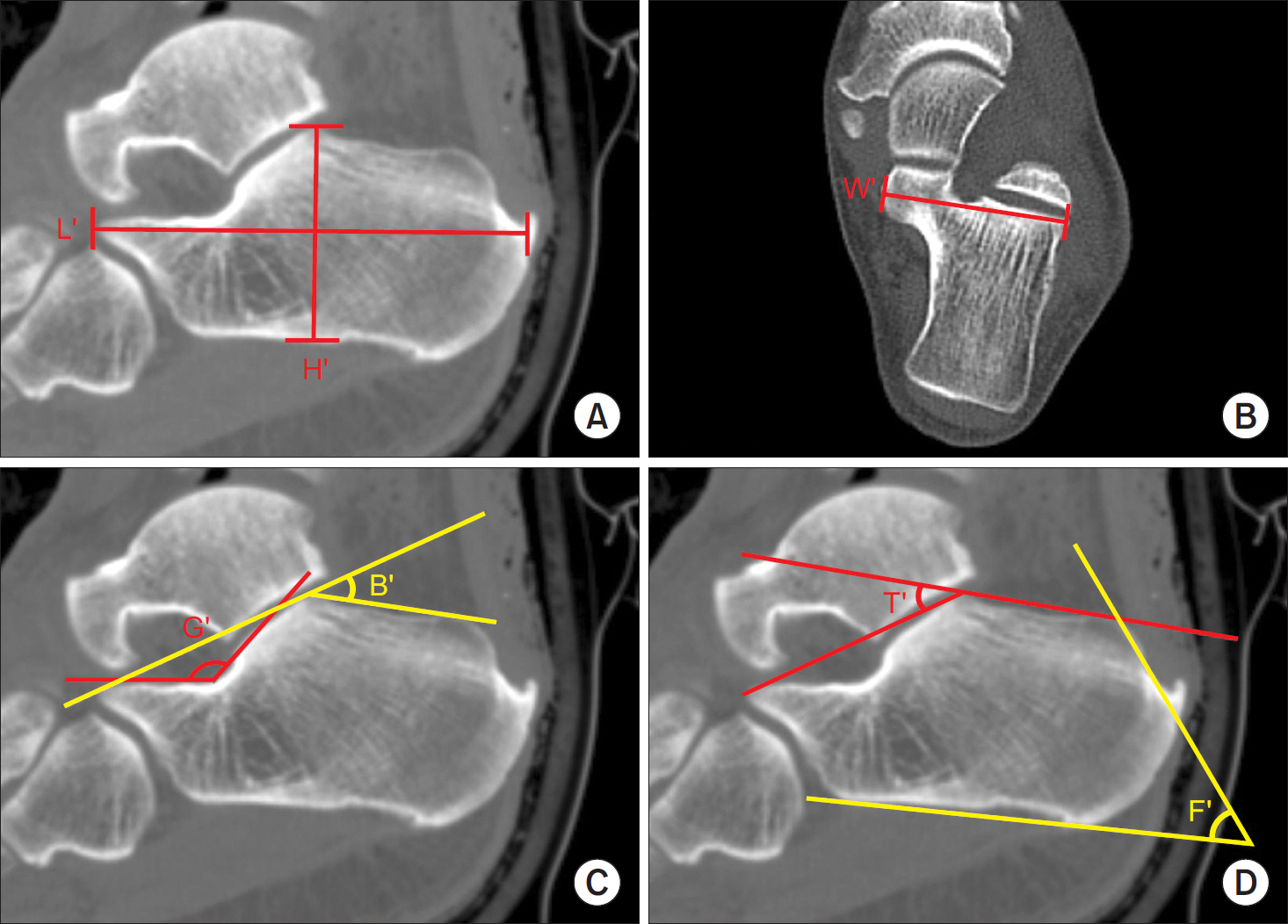
Figure 3. Radiologic measurement of calcaneus using computed tomography images. (A) L': maximum length of calcaneus, H': maximum height of calcaneus. (B) W': maximal width of calcaneus. (C) B': Böhler angle, G': Gissane angle. (D) T': tala-articular angle, F': Fowler-Philip angle.
Reference
-
References
1. Koshy S, Vettivel S, Selvaraj KG. Estimation of length of calcaneum and talus from their bony markers. Forensic Sci Int. 2002; 129:200–4.
Article2. Smith SL. Stature estimation of 3–10-year-old children from long bone lengths. J Forensic Sci. 2007; 52:538–46.
Article3. Brough AL, Bennett J, Morgan B, Black S, Rutty GN. Anthropological measurement of the juvenile clavicle using multidetector computed tomography–affirming reliability. J Forensic Sci. 2013; 58:946–51.
Article4. Hildebolt CF, Vannier MW, Knapp RH. Validation study of skull three-dimensional computerized tomography measurements. Am J Phys Anthropol. 1990; 82:283–94.
Article5. Lopes PM, Moreira CR, Perrella A, Antunes JL, Cavalcanti MG. 3-D volume rendering maxillofacial analysis of angular measurements by multislice CT Oral Surg Oral Med Oral Pathol Oral Radiol Endod. 2008; 105:224–30.6. Robinson C, Eisma R, Morgan B, Jeffery A, Graham EA, Black S, et al. Anthropological measurement of lower limb and foot bones using multidetector computed tomography. J Forensic Sci. 2008; 53:1289–95.
Article7. Verhoff MA, Ramsthaler F, Krähahn J, Deml U, Gille RJ, Grabherr S, et al. Digital forensic osteology–possibilities in cooperation with the Virtopsy project. Forensic Sci Int. 2008; 174:152–6.
Article8. Guyomarc'h P, Santos F, Dutailly B, Desbarats P, Bou C, Co-queugniot H. Three-dimensional computer-assisted craniomet-rics: a comparison of the uncertainty in measurement induced by surface reconstruction performed by two computer programs. Forensic Sci Int. 2012; 219:221–7.9. Sholts SB, Flores L, Walker PL, Wärmländer SKTS. Comparison of coordinate measurement precision of different landmark types on human crania using a 3D laser scanner and a 3D digitiser: Implications for applications of digital morphometrics. Int J Osteoarchaeol. 2011; 21:535–43.
Article
