Primary Renal Undifferentiated Sarcoma as an Infiltrative Mass in a 12-Year-Old Boy
- Affiliations
-
- 1Department of Radiology and Research Institute of Radiological Science, Severance Children's Hospital, Yonsei University College of Medicine, Seoul, Korea. mjl1213@yuhs.ac
- 2Department of Pathology, Severance Hospital, Yonsei University College of Medicine, Seoul, Korea.
- KMID: 2043613
- DOI: http://doi.org/10.3348/jksr.2015.73.3.199
Abstract
- Undifferentiated sarcomas are rare tumors not classified into any sarcoma subtype. Due to their rarity, imaging findings of undifferentiated sarcomas are poorly characterized. The purpose of this report was to present imaging findings of a pathologically confirmed undifferentiated sarcoma originated from the left kidney of a 12-year-old boy. The mass was infiltrative involving the renal pelvis. It mimicked massive hilar lymphadenopathy with a preserved renal contour visible by both ultrasonography and CT. Renal vein thrombosis was also observed. Although undifferentiated sarcomas are rare, they should be considered in differential diagnosis of infiltrative renal masses with renal pelvis invasion in children.
MeSH Terms
Figure
-
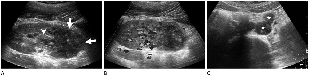
Fig. 1 Ultrasonography of a 12-year-old boy presented with recurrent abdominal pain and vomiting. A. Abdominal ultrasonography with longitudinal scan of the left kidney showing an ill-defined and heterogeneously hypoechoic mass (arrows) within the mid to lower pole of the kidney with a preserved renal contour. The mass seems to invade the renal hilum. It is accompanied by pelvic wall thickening (arrowhead). B. Doppler interrogation of the left kidney revealing some vascularity within the mass with much less vascularity compared to the adjacent normal renal parenchyma. C. Ultrasonography with transverse scan of the mid-abdomen just below the level of the left renal vein showing hypoechoic nodular lesions (asterisks) considered as enlarged lymph nodes in the left paraaortic area.
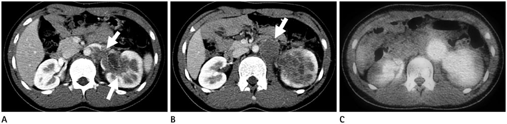
Fig. 2 Further imaging studies of a 12-year-old boy with left renal mass. A. Abdomino-pelvic computed tomography (CT) of an axial image of the nephrographic phase at the level of the left renal hilum demonstrating an infiltrative renal mass involving the renal pelvis with low-attenuating lesions (arrows) within the expected course of the left renal vein, suggestive of venous thrombosis. B. An axial slice of CT slightly caudad to A showing an ill-defined and low attenuating lesion with heterogeneous enhancement replacing the left renal parenchyma with a preserved renal contour. A round low-attenuation lesion (arrow) between the aorta and the left renal tumor is also noted. This lesion was thought to represent metastatic lymphadenopathy at the time of diagnosis. However, it was later confirmed to be a massive renal hilar and perirenal extension of the tumor. C. Axial fusion positron emission tomography-CT scan taken on approximately the same level as B showing intense fluorodeoxyglucose uptake in both the left renal and perihilar masses.
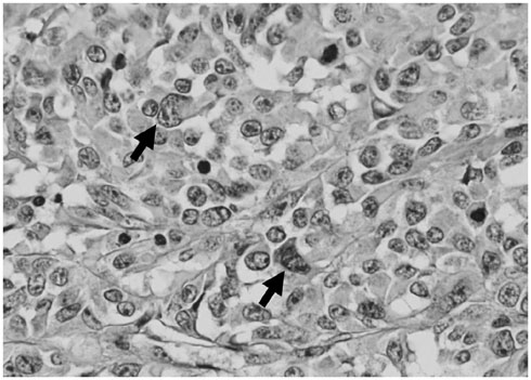
Fig. 3 Microscopy demonstrating anaplastic round to polygonal tumor cells (arrows) with vesicular nuclei and prominent nucleoli (hematoxylin and eosin, original magnification × 400). The tumor cells show abundant cytoplasm and high mitotic rate.
Reference
-
1. Lowe LH, Isuani BH, Heller RM, Stein SM, Johnson JE, Navarro OM, et al. Pediatric renal masses: Wilms tumor and beyond. Radiographics. 2000; 20:1585–1603.2. Lalwani N, Prasad SR, Vikram R, Katabathina V, Shanbhogue A, Restrepo C. Pediatric and adult primary sarcomas of the kidney: a cross-sectional imaging review. Acta Radiol. 2011; 52:448–457.3. Alaggio R, Bisogno G, Rosato A, Ninfo V, Coffin CM. Undifferentiated sarcoma: does it exist? A clinicopathologic study of 7 pediatric cases and review of literature. Hum Pathol. 2009; 40:1600–1610.4. Stein-Wexler R. Pediatric soft tissue sarcomas. Semin Ultrasound CT MR. 2011; 32:470–488.5. Siegel MJ, Chung EM. Wilms' tumor and other pediatric renal masses. Magn Reson Imaging Clin N Am. 2008; 16:479–497. vi6. Downey RT, Dillman JR, Ladino-Torres MF, McHugh JB, Ehrlich PF, Strouse PJ. CT and MRI appearances and radiologic staging of pediatric renal cell carcinoma. Pediatr Radiol. 2012; 42:410–417. quiz 513-5147. Chepuri NB, Strouse PJ, Yanik GA. CT of renal lymphoma in children. AJR Am J Roentgenol. 2003; 180:429–431.8. Raney B, Anderson J, Arndt C, Crist W, Maurer H, Qualman S, et al. Primary renal sarcomas in the Intergroup Rhabdomyosarcoma Study Group (IRSG) experience, 1972-2005: a report from the Children's Oncology Group. Pediatr Blood Cancer. 2008; 51:339–343.9. Somers GR, Gupta AA, Doria AS, Ho M, Pereira C, Shago M, et al. Pediatric undifferentiated sarcoma of the soft tissues: a clinicopathologic study. Pediatr Dev Pathol. 2006; 9:132–142.
- Full Text Links
-
- Actions
-
Cited
- CITED
-
- Close
- Share
-
- Similar articles
-
- Primary renal BCOR::CCNB3 sarcoma in a female patient: case report
- Cutaneous Metastatic Undifferentiated Pleomorphic Sarcoma from a Mediastinal Sarcoma
- Primary Undifferentiated Pleomorphic Sarcoma of the Colon Mesentery
- Two cases of primary renal sarcoma
- Undifferentiated ( Embryonal ) Sarcoma of the Liver: A case report including immunohistochemical, electronmicroscopic and flow-cytometric study
