A Case Report of Localized Hepatic Sinusoidal Dilatation: The Diagnostic Usefulness of the Hepatobiliary Phase of Gd-EOB-DTPA-Enhanced Magnetic Resonance Imaging
- Affiliations
-
- 1Department of Radiology, Daegu Fatima Hospital, Daegu, Korea. nosmokeman@naver.com
- KMID: 2043610
- DOI: http://doi.org/10.3348/jksr.2015.73.3.186
Abstract
- Hepatic sinusoidal dilatation (HSD) is a rare vascular disorder characterized by focal dilatation of the sinusoidal spaces in the liver. In most cases, it may be associated with venous outflow impairment. In addition, this histological change could occur in a number of systemic and hepatic conditions in the absence of hepatic venous obstruction. However, the pathogenesis has not yet been elucidated. To the best of our knowledge, imaging findings in a case of localized HSD without any additional medical disorder or oral contraceptive therapy have not been described previously in the literature written in English. Here, we describe imaging findings in a case of localized HSD mimicking a hepatic tumor, focusing on the useful findings on the gadolinium ethoxybenzyl diethylenetriamine pentaacetic acid-enhanced hepatobiliary phase MR image.
Figure
-
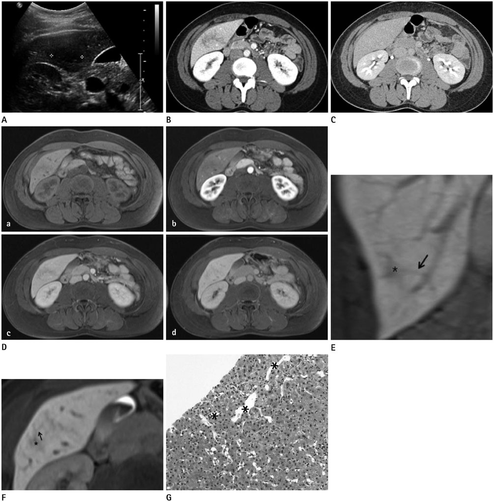
Fig. 1 A 37-year-old woman with localized hepatic sinusoidal dilatation (HSD). A. Transabdominal ultrasonography showing an ill-defined and hypoechoic lesion (approximately 3 cm) in hepatic segment VI. B. The axial enhanced CT image during the portal venous phase (PVP) (60 sec) showing an ill-defined and hypodense structure with an almost isodense portion in hepatic segment VI. C. The axial enhanced CT image during the equilibrium phase (3 min) showing the lesion as isodense compared with adjacent liver parenchyma. D. Contrast-enhanced dynamic MR HSD images during arterial, portal venous, and transitional phases, including an unenhanced T1-weighted image. a. On the unenhanced T1-weighted image, a focal, ill-defined, slightly hypointense hepatic lesion is noted. b. In the arterial phase, the lesion is indistinct. c. In the PVP, an ill-defined and hypointense structure with an almost isointense area, as compared with adjacent liver parenchyma, is observed. d. In the transitional phase, the lesion becomes less prominent than in the PVP. E, F. On the axial (E) and reformatted coronal (F) MR images of the delayed hepatobiliary phase, the ill-defined, vessel-like hypointense structure (asterisk) is connected to the peripheral branch of the right portal vein and the middle hepatic vein, respectively (black arrow). G. Photomicrograph showing the only sinusoidal dilatation (asterisk) without peliosis in the liver tissue obtained by needle biopsy (hematoxylin and eosin, × 200).
Reference
-
1. Bruguera M, Caballero T, Carreras E, Aymerich M, Rodés J, Rozman C. Hepatic sinusoidal dilatation in Hodgkin's disease. Liver. 1987; 7:76–80.2. Capron JP, Lemay JL, Gontier MF, Dupas JL, Capron-Chivrac D, Lorriaux A. Hepatic sinusoidal dilatation in Crohn's disease. Scand J Gastroenterol. 1979; 14:987–992.3. Fisher MR, Neiman HL. Periportal sinusoidal dilatation associated with pregnancy. Cardiovasc Intervent Radiol. 1984; 7:299–302.4. Rubbia-Brandt L, Audard V, Sartoretti P, Roth AD, Brezault C, Le Charpentier M, et al. Severe hepatic sinusoidal obstruction associated with oxaliplatin-based chemotherapy in patients with metastatic colorectal cancer. Ann Oncol. 2004; 15:460–466.5. Bruguera M, Aranguibel F, Ros E, Rodés J. Incidence and clinical significance of sinusoidal dilatation in liver biopsies. Gastroenterology. 1978; 75:474–478.6. Weinberger M, Garty M, Cohen M, Russo Y, Rosenfeld JB. Ultrasonography in the diagnosis and follow-up of hepatic sinusoidal dilatation. Arch Intern Med. 1985; 145:927–929.7. Yang DM, Jung DH, Park CH, Kim JE, Choi SJ. Imaging findings of hepatic sinusoidal dilatation. AJR Am J Roentgenol. 2004; 183:1075–1077.8. Apicella PL, Mirowitz SA, Weinreb JC. Extension of vessels through hepatic neoplasms: MR and CT findings. Radiology. 1994; 191:135–136.9. Shin NY, Kim MJ, Lim JS, Park MS, Chung YE, Choi JY, et al. Accuracy of gadoxetic acid-enhanced magnetic resonance imaging for the diagnosis of sinusoidal obstruction syndrome in patients with chemotherapy-treated colorectal liver metastases. Eur Radiol. 2012; 22:864–871.10. Gouya H, Vignaux O, Legmann P, de Pigneux G, Bonnin A. Peliosis hepatis: triphasic helical CT and dynamic MRI findings. Abdom Imaging. 2001; 26:507–509.
- Full Text Links
-
- Actions
-
Cited
- CITED
-
- Close
- Share
-
- Similar articles
-
- Hepatic Lymphoma Representing Iso-Signal Intensity on Hepatobiliary Phase, in Gd-EOB-DTPA-Enhanced MRI: Case Report
- Image Findings of Sarcomatous Intrahepatic Cholangiocarcinoma Focused on Gd-EOB-DTPA Enhanced MRI: A Case Report
- Supradiaphragmatic Liver Confirmed by a Hepatocyte-specific Contrast Agent (Gd-EOB-DTPA): A Case Report
- Confident Diagnosis of Bronchobiliary Fistula Using Contrast-Enhanced Magnetic Resonance Cholangiography
- Enhancement Pattern of Liver Parenchyma during Late Dynamic Phase Imaging: Comparison between Gd-EOB-DTPA and Gd-DTPA-BMA
