Congenital Hypoplasia of the Medial Hallucial Sesamoid with Avascular Necrosis: A Case Report
- Affiliations
-
- 1Department of Radiology, Hanyang University College of Medicine, Seoul Hospital, Seoul, Korea. radsh@medimail.co.kr
- 2Department of Rheumatology, Hanyang University College of Medicine, Seoul Hospital, Seoul, Korea.
- 3Department of Radiology, Hanyang University College of Medicine, Guri Hospital, Guri, Korea.
- 4Department of Pathology, National Police Hospital, Seoul, Korea.
- KMID: 2041953
- DOI: http://doi.org/10.3348/jksr.2013.69.4.311
Abstract
- Avascular necrosis of the hallucial sesamoids is an uncommon cause of metatarsalgia, and the congenital absence of the medial sesamoid is also a rarely reported condition in the podiatric literature. It must be distinguished from other painful conditions of the sesamoid due to the opposite direction of treatment. To our knowledge, there is no reported case of congenital hypoplasia of the medial sesamoid with osteonecrosis. We report a case of nontraumatic metatarsal pains with progressive sclerosis and fragmentation of the medial sesamoid on serial radiographs, magnetic resonance imaging, and ultrasonography with an incidental finding for the absence of contralateral medial sesamoid in a 33-year-old female.
MeSH Terms
Figure
-

Fig. 1 Plain radiographs of the right foot who a 33-year-old woman with plantar pain to the right first metatarsophalangeal joint. A comparison of two radiographs at interval changes of a year; the medial sesamoid collapsed and had more areas of demineralization with fragmentation. A. Initial right lateral ankle radiograph at the outside hospital demonstrates flattening of the medial sesamoid with sclerosis (arrow). B. A year after initial lateral radiograph demonstrates a more collapsed medial sesamoid with increased areas of demineralization (arrow). C. An additional sesamoid view demonstrates the absence of the left medial sesamoid (arrows) and hypoplasia of the right medial sesamoid (arrows) with fragmentation and adjacent soft tissue swelling.

Fig. 2 Ultrasonography of the right foot. A. A transverse scan shows multiple echogenic nodules of the medial sesamoid (arrow) with fragmentation. B. A longitudinal scan shows an irregular surface of the medial sesamoid (arrow) with multiple echogenic nodules.
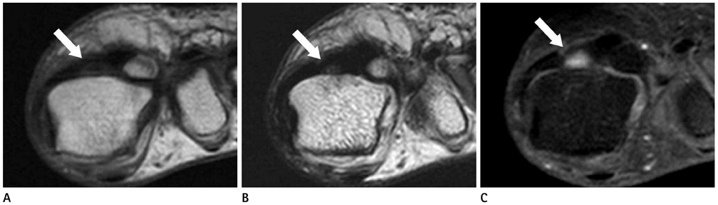
Fig. 3 Magnetic resonance image (MRI) of the right foot. A. T1-weighted coronal MR image demonstrates the low signal intensity of the right medial sesamoid (arrow). B. T2-weighted coronal MR image demonstrates heterogenously low to intermediate signal intensity with fragementation of the right medial sesamoid (arrow). C. On gadolinium enhanced, fat-suppressed T1-weighted coronal MR image demonstrates patchy localized signal enhancement seen within the medial sesamoid (arrow). These findings reflect avascular necrosis of the medial sesamoid.
Reference
-
1. Karasick D, Schweitzer ME. Disorders of the hallux sesamoid complex: MR features. Skeletal Radiol. 1998; 27:411–418.2. Bodner G, Stöckl B, Fierlinger A, Schocke M, Bernathova M. Sonographic findings in stress fractures of the lower limb: preliminary findings. Eur Radiol. 2005; 15:356–359.3. Sanders TG, Rathur SK. Imaging of painful conditions of the hallucal sesamoid complex and plantar capsular structures of the first metatarsophalangeal joint. Radiol Clin North Am. 2008; 46:1079–1092. vii4. Lafforgue P. Pathophysiology and natural history of avascular necrosis of bone. Joint Bone Spine. 2006; 73:500–507.5. Toussirot E, Jeunet L, Michel F, Kantelip B, Wendling D. Avascular necrosis of the hallucal sesamoids update with reference to two case-reports. Joint Bone Spine. 2003; 70:307–309.6. Coughlin MJ. Sesamoids and Accessory Bones of the Foot. In : Roger AM, editor. Surgery of the foot and ankle. 8th ed. Philadelphia, PA: Mosby;2007. p. 531–543.7. Kanatli U, Ozturk AM, Ercan NG, Ozalay M, Daglar B, Yetkin H. Absence of the medial sesamoid bone associated with metatarsophalangeal pain. Clin Anat. 2006; 19:634–639.8. Taylor JA, Sartoris DJ, Huang GS, Resnick DL. Painful conditions affecting the first metatarsal sesamoid bones. Radiographics. 1993; 13:817–830.9. Jahss MH. The sesamoids of the hallux. Clin Orthop Relat Res. 1981; 88–97.10. Scranton PE Jr, Rutkowski R. Anatomic variations in the first ray: Part II. Disorders of the sesamoids. Clin Orthop Relat Res. 1980; 256–264.
- Full Text Links
-
- Actions
-
Cited
- CITED
-
- Close
- Share
-
- Similar articles
-
- Avascular Necrosis of the Hallucal Sesamoid: Three Cases Report
- Gout of the Hallucal Medial Sesamoid: A Case Report
- A Study on the Changes of the Femoral Head following Treatment of Congenital Dislocation of the Hip
- The Results of Distal Chevron Osteotomy with Lateral Soft Tissue Release for Hallux Valgus Deformity
- Bilateral Hypoplasia of the Medial and Lateral Menisci
