Korean J Obstet Gynecol.
2011 Feb;54(2):103-106. 10.5468/KJOG.2011.54.2.103.
An ectopic ureterocele diagnosed in interlabial mass
- Affiliations
-
- 1Department of Obstetrics and Gynecology, Keimyung University School of Medicine, Daegu, Korea. jcpark@dsmc.or.kr
- 2Department of Urology, Keimyung University School of Medicine, Daegu, Korea.
- KMID: 2037499
- DOI: http://doi.org/10.5468/KJOG.2011.54.2.103
Abstract
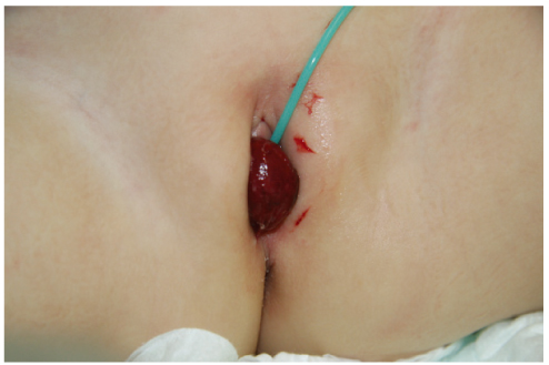
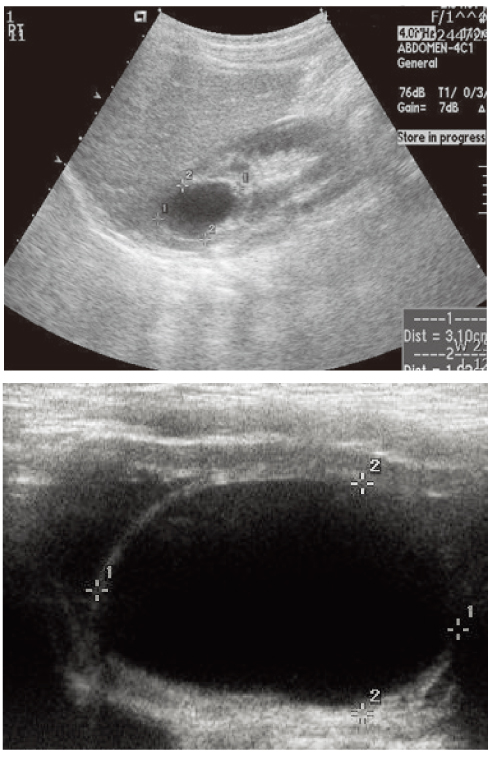
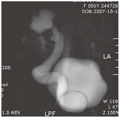
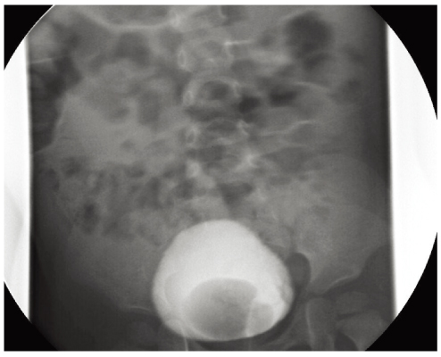
- Interlabial mass in infant is not common. Because of similarity of symptoms and signs of those mass and less experience of gynecologist due to those rarity, differential diagnosis is not easy. In infant, there are five common interlabial masses which are prolapsed ectopic ureterocele, urethral prolapse, paraurethral cyst, hydrocolpos and rhabdomyosarcoma. Ureterocele with duplex ureter might be diagnosed, however prolapsed ureterocele through urethra is extremely rare. We found 18 months old girl with interlabial mass and diagnosed as prolapsed ectopic ureterocele by ultrasonography, magnetic resonance imaging and voiding cystourethrography. We managed by endoscopic incision of ureterocele successfully. So we report this case with a brief review of associated literatures.
Keyword
MeSH Terms
Figure
Reference
-
1. Fletcher SG, Lemack GE. Benign masses of the female periurethral tissues and anterior vaginal wall. Curr Urol Rep. 2008. 9:389–396.2. Nussbaum AR, Lebowitz RL. Interlabial masses in little girls: review and imaging recommendations. AJR Am J Roentgenol. 1983. 141:65–71.3. Seo CS, Lee MH, Hwang ES, Kim SH, Kim HS, Lee KW. A case of bilateral duplex ureter with ureteroceles diagnosed by prenatal ultrasound. Korean J Obstet Gynecol. 1994. 37:1858–1864.4. Scott JE. The single ectopic ureter and the dysplastic kidney. Br J Urol. 1981. 53:300–305.5. Klauber GT, Crawford DB. Prolapse of ectopic ureterocele and bladder trigone. Urology. 1980. 15:164–166.6. Moffett JD, Banks R Jr. Prolapse of the urethra in young girls. J Am Med Assoc. 1951. 146:1288–1290.7. Devine PC, Kessel HC. Surgical correction of urethral prolapse. J Urol. 1980. 123:856–857.8. Blaivas JG, Pais VM, Retik AB. Paraurethral cysts in female neonate. Urology. 1976. 7:504–507.9. Das SP. Paraurethral cysts in women. J Urol. 1981. 126:41–43.10. Hahn-Pedersen J, Kvist N, Nielsen OH. Hydrometrocolpos: current views on pathogenesis and management. J Urol. 1984. 132:537–540.11. Flamant F, Gerbaulet A, Nihoul-Fekete C, Valteau-Couanet D, Chassagne D, Lemerle J. Long-term sequelae of conservative treatment by surgery, brachytherapy, and chemotherapy for vulval and vaginal rhabdomyosarcoma in children. J Clin Oncol. 1990. 8:1847–1853.12. Mierau GW, Favara BE. Rhabdomyosarcoma in children: ultrastructural study of 31 cases. Cancer. 1980. 46:2035–2040.13. Arap S, Nahas WC, Alonso G, Denes FT, Martins LR, Menezes de Goes G. Assessment of hydroureteronephrosis by renographic evaluation under diuretic stimulus. Urol Int. 1984. 39:170–174.14. Ilica AT, Kocaoglu M, Bulakbasi N, Surer I, Tayfun C. Prolapsing ectopic ureterocele presenting as a vulval mass in a newborn girl. Diagn Interv Radiol. 2008. 14:33–34.15. Coplen DE. Management of the neonatal ureterocele. Curr Urol Rep. 2001. 2:102–105.
- Full Text Links
- Actions
-
Cited
- CITED
-
- Close
- Share
- Similar articles
-
- An ectopic ureterocele presented as a prolapsed vaginal mass in a 1-month-old girl
- A Case of Ectopic Ureterocele Associated with Reflux into the Ureterocele
- A Case of Giant Hydronephrosis with Ectopic Ureterocele in Bilateral Duplicated Ureters
- Ectopic ureterocele in children
- Two Cases of Simple Ureterocele


