An Intratendinous Tophaceous Gout Mistaken for Cellulitis in the Patellar Tendon
- Affiliations
-
- 1Department of Orthopaedic Surgery, National Police Hospital, Seoul, Korea. jdoc3846@google.com
- KMID: 2035872
- DOI: http://doi.org/10.4055/jkoa.2015.50.4.337
Abstract
- Gout is characterized by recurrent attacks of arthralgia, and deposition of monosodium urate crystals in and around the joints of the extremities and soft tissues. Monosodium urate crystals are observed most frequently at the 1st metatarsophalangeal joint and usually presented in the ankle and wrist joint. However, no case of an intratendinous tophus in the patellar tendon has been reported in Korean literature. In this report, we found monosodium urate crystals in the patellar tendon on magnetic resonance imaging images and intratendinous tophus were visible to the naked eye by excision. We reported on the case of a patient who experienced an unusual intratendinous tophus in the patellar tendon.
Keyword
MeSH Terms
Figure
-

Figure 1 Knee joint anterioposterior view (A) and knee joint lateral view (B) shows no definite findings.
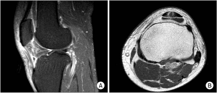
Figure 2 (A) T1-weighted spiral sagittal enhancement magnetic resonance imaging (MRI) shows edematous change from the prepatellar area to the pretibial area and rim enhancement at the infrapatellar fat pad area. (B) T1-weighted axial enhancement MRI shows fluid collection and peripheral rim enhancement in the middle of the patellar and retro-patellar tendon area.
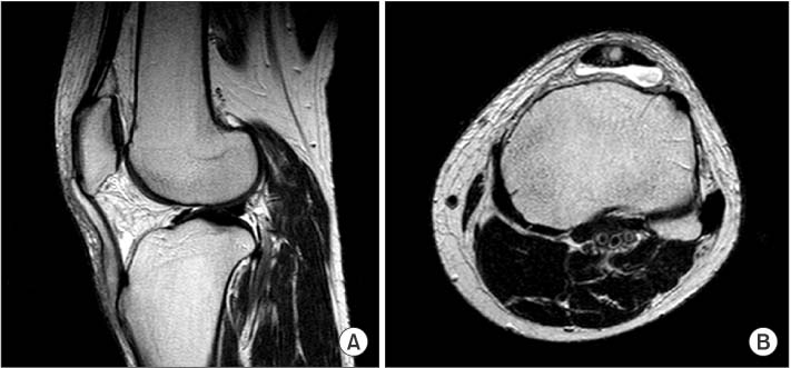
Figure 3 (A) T2-weighted sagittal magnetic resonance imaging (MRI) shows intratendinous fluid collection along the patellar tendon and infrapatellar fat pad area. (B) T2-weighted axial MRI shows fluid collection in the middle of the patellar and retro-patellar tendon area.

Figure 4 (A) Intraoperative photograph shows white caseous materials. (B) Below the white caseous material, yellowish fluid collection was found.

Figure 5 Chalky white mass was found after dissection.
Reference
-
1. Kim MK, Moon KH, Lee TJ, Kim L, Lee JS. Gout tophi in the bipartite patella: a case report. J Korean Orthop Assoc. 2008; 43:139–142.2. Gililland JM, Webber NP, Jones KB, Randall RL, Aoki SK. Intratendinous tophaceous gout imitating patellar tendonitis in an athletic man. Orthopedics. 2011; 34:223.
Article3. Gerster JC, Landry M, Rappoport G, Rivier G, Duvoisin B, Schnyder P. Enthesopathy and tendinopathy in gout: computed tomographic assessment. Ann Rheum Dis. 1996; 55:921–923.
Article4. Loeb JN. The influence of temperature on the solubility of monosodium urate. Arthritis Rheum. 1972; 15:189–192.
Article5. Straub LR, Smith JW, Carpenter GK Jr, Dietz GH. The surgery of gout in the upper extremity. J Bone Joint Surg Am. 1961; 43:731–774.
Article6. Narváez JA, Narváez J, Ortega R, De Lama E, Roca Y, Vidal N. Hypointense synovial lesions on T2-weighted images: differential diagnosis with pathologic correlation. AJR Am J Roentgenol. 2003; 181:761–769.7. Jennings F, Lambert E, Fredericson M. Rheumatic diseases presenting as sports-related injuries. Sports Med. 2008; 38:917–930.
Article8. Beskin JL, Sanders RA, Hunter SC, Hughston JC. Surgical repair of Achilles tendon ruptures. Am J Sports Med. 1987; 15:1–8.
Article9. Tashiro S, Sugita T, Nakamura S, Kurata Y. Gout tophus in the bipartite patella. Orthopedics. 2002; 25:1295–1296.
Article10. Levy M, Seelenfreund M, Maor P, Fried A, Lurie M. Bilateral spontaneous and simultaneous rupture of the quadriceps tendons in gout. J Bone Joint Surg Br. 1971; 53:510–513.
Article
- Full Text Links
-
- Actions
-
Cited
- CITED
-
- Close
- Share
-
- Similar articles
-
- Intratendinous Tophaceous Gout Mimicking Cellulitis after Achilles Tendon Repair
- Acute Tophaceous Gout of the Distal Interphalangeal Joint Misdiagnosed as Cellulitis
- Complete Rupture of the Tibialis Anterior Tendon Due to Intratendinous Ganglion Cyst
- Tophaceous Gout Involving the Bipartitle Patella: A Case Report
- Tophaceous Gout in the Rotator Cuff with Impingement Syndrome: A Case Report
