Treatment of Delayed Cerebrospinal Fluid Rhinorrhea after Head Trauma: A Case Report
- Affiliations
-
- 1Department of Neurological Surgery, The Armed Forced Capital Hospital, Seongnam, Korea. nscharisma@hanmail.net
- KMID: 2019932
- DOI: http://doi.org/10.13004/jknts.2009.5.1.33
Abstract
- Most of cerebrospinal fluid (CSF) leakages after head trauma are presented within several days and improved by conservative management, although it often needs special procedure, such as lumbar drainage. However, sometimes surgeries are required if patients do not improve by non-surgical treatments. We have experienced one case of 20 year-old man undertaken two operations because of head trauma and nasal CSF leakage, which was presented 29 days after trauma and had not been cured by conservative management. In the operating field, we found the fistula from anterior fossa to nasal cavity. In order to seal the fistula, we used galea flap. We report this case with literature reviews.
Figure
-
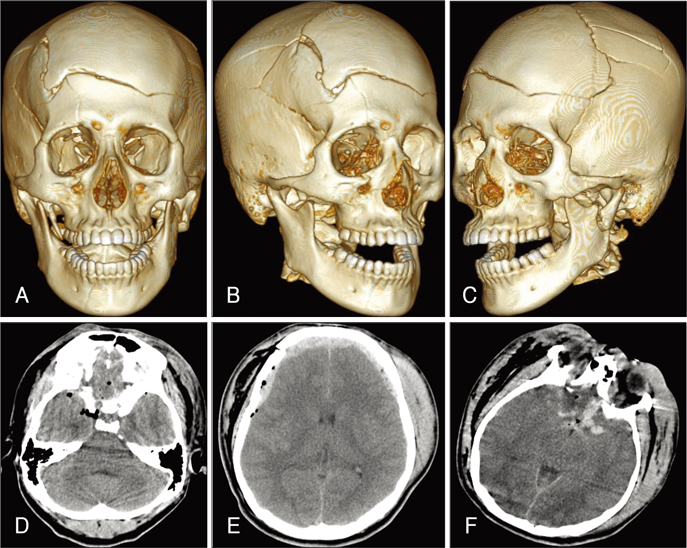
FIGURE 1 PreOP images. AC: Multiple skull fractures, basal skull fractures and facial bone fractures were shown on 3-dimensional computed tomography (CT) reconstruction. D, E: Brain CT demonstrated forehead swelling with pneumocephalus and epidural hematoma on the bifrontal areas. F: 3 hours after trauma hemorrhagic contusion was shown on frontal base.
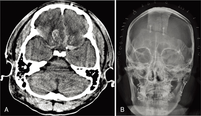
FIGURE 2 PostOP images. A, B: After bilateral decompressive craniectomy, hemorrhagic contusion and swelling of both frontal lobes were shown on the CT.
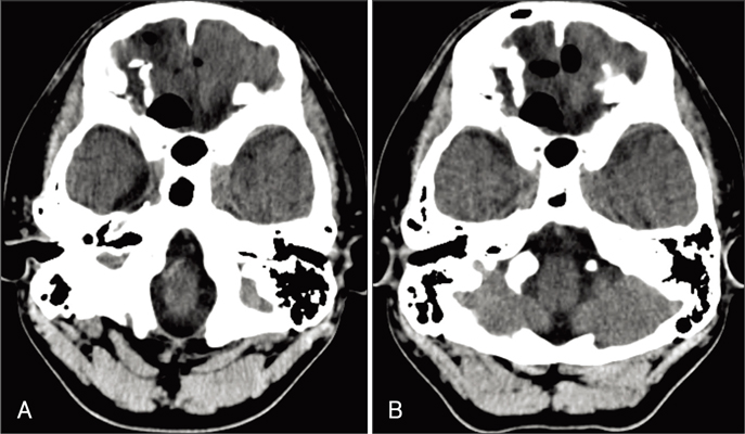
FIGURE 3 CT images with CSF rhinorrhea. A: Brain CT scans taken 29 days after trauma show pneumocephalus on the frontal base. B: CT taken 55 days after trauma shows no interval change of pneumocephalus.
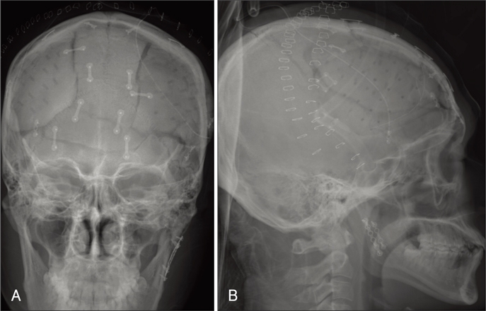
FIGURE 4 PostOP images. A, B: Skull anteriorposterior (AP) and lateral view of x-ray after reconstruction surgery with galeal frontalis myofascial flap and cranioplasty.
Reference
-
1. Eljamel MS, Foy PM. Post-traumatic CSF fistulae, the case for surgical repair. Br J Neurosurg. 1990; 4:479–483.
Article2. Friedman JA, Ebersold MJ, Quast LM. Post-traumatic cerebrospinal fluid leakage. World J Surg. 2001; 25:1062–1066.
Article3. Kim HS, Hur JW, Lee JW, Lee HK. Extraordinarily long-term posttraumatic cerebrospinal fluid fistula. J Korean Neurosurg Soc. 2007; 42:403–405.
Article4. Martin TJ, Loehrl TA. Endoscopic CSF leak repair. Curr Opin Otolaryngol Head Neck Surg. 2007; 15:35–39.
Article5. Moza K, McMenomey SO, Delashaw JB Jr. Indications for cerebrospinal fluid drainage and avoidance of complications. Otolaryngol Clin North Am. 2005; 38:577–582.
Article6. Schick B, Weber R, Kahle G, Draf W, Lackmann GM. Late manifestations of traumatic lesions of the anterior skull base. Skull Base Surg. 1997; 7:77–83.
Article7. Scholsem M, Scholtes F, Collignon F, Robe P, Dubuisson A, Kaschten B, et al. Surgical management of anterior cranial base fractures with cerebrospinal fluid fistulae: a single-institution experience. Neurosurgery. 2008; 62:463–469. discussion 469-471.8. Shapiro SA, Scully T. Closed continuous drainage of cerebrospinal fluid via a lumbar subarachnoid catheter for treatment or prevention of cranial/spinal cerebrospinal fluid fistula. Neurosurgery. 1992; 30:241–245.
Article9. Stewart BT, Kaye AH. Delayed cerebrospinal fluid rhinorrhoea: a case report. Aust N Z J Surg. 1992; 62:818–820.
Article
- Full Text Links
-
- Actions
-
Cited
- CITED
-
- Close
- Share
-
- Similar articles
-
- Endoscopic Repair of Spontaneous Cerebrospinal Fluid Rhinorrhea with a Nasoseptal Flap
- Endoscopic treatment of iatrogenic cerebrospinal fluid rhinorrhea
- Management of Cerebrospinal Fluid Rhinorrhea
- Surgical management of iatrogenic cerebrospinal fluid rhinorrhea
- Traumatic Cerebrospinal Fluid Rhinorrhea: Successful Closure under the Surgical Microscope
