Granulocytic Sarcoma in the Head and Neck: CT and MR Imaging Findings
- Affiliations
-
- 1Department of Radiology, College of Medicine, Inha University, Incheon, Korea. swpark88@inha.ac.kr
- 2Department of Radiology, College of Medicine, Seoul National University, Seoul, Korea.
- 3Department of Radiology, Samsung Medical Center, Sungkyunkwan University School of Medicine, Seoul, Korea.
- KMID: 2007187
- DOI: http://doi.org/10.3342/ceo.2009.2.2.66
Abstract
OBJECTIVES
To evaluate characteristic computed tomography (CT) and magnetic resonance (MR) imaging findings of granulocytic sarcomas of the head and neck.
METHODS
The CT (n=11) and MR (n=1) images obtained from 11 patients (7 males and 4 females; mean age, 23.5 yr; age range, 1 to 69 yr) with histologically-proven granulocytic sarcomas of the head and neck were retrospectively reviewed. Histological confirmation was done by bone marrow biopsy in 9 patients, and/or local biopsy in 4 patients. The imaging findings were analyzed with particular attention to location, size, shape, margin, bone destruction, internal architecture, pattern and degree of enhancement, and multiplicity of the lesions.
RESULTS
The masses were most commonly located in the orbital cavity (n=8); other locations included lymph nodes (n=5) and palatine/pharyngeal/lingual tonsils (n=3). The mass sizes varied from a mean diameter of 1.3 to 5.8 cm (average, 2.6 cm). Multiple lesions were found in 6 patients. The shapes of the tumors were ovoid in 12 patients and irregular in 4 patients. Most lesions had poorly-defined margins (13/16) and invaded adjacent bony structures (5/16). On the pre-contrast CT images, the masses were iso- (5/8) or low-density (3/8) in comparison with muscle. The MRI, which was obtained in one patient in this study, showed that the mass was iso-signal intensity on T1-weighted images and iso-signal intensity on T2-weighted images compared to the gray matter of the brain. On the post-contrast CT images, there was homogenesous (n=12) or heterogeneous (n=4) enhancement, with mild (n=10), moderate (n=4), and marked (n=2) enhancement in the solid portions of the lesions.
CONCLUSION
Although rare, granulocytic sarcomas arise in various locations in the head and neck area (most commonly in the orbit) in the form of well-demarcated, and mildly- and homogenously-enhancing masses with adjacent bony invasion.
Keyword
MeSH Terms
Figure
-
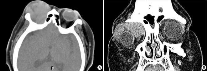
Fig. 1 A 27-yr-old woman with a 1-yr history of acute myeloid leukemia presented with painful, reddening of the right orbit. (A) Post-contrast axial CT image shows homogeneously-enhancing soft tissue mass with irregular shape in intraconal and extraconal spaces of the right orbit. (B) Post-contrast coronal CT image shows large enhancing mass without adjacent bone involvement in the right orbit.
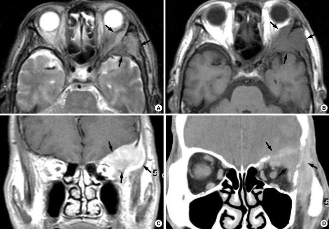
Fig. 2 A 69-yr-old man with a 7-yr history of chronic myeloid leukemia presented with left orbital pain. (A) T2-weighted axial image shows an iso-signal intensity mass (arrows) in the lateral side of left orbit. (B) T1-weighted axial image shows an iso-signal intensity mass (arrows). Adjacent bone marrow was involved by the tumor. (C) The mass is well-enhanced on the post-contrast T1-weighted coronal MR image (arrows). (D) Post-contrast coronal CT image shows the mass (arrows) involving the adjacent sphenoid bone and the intracranial area.
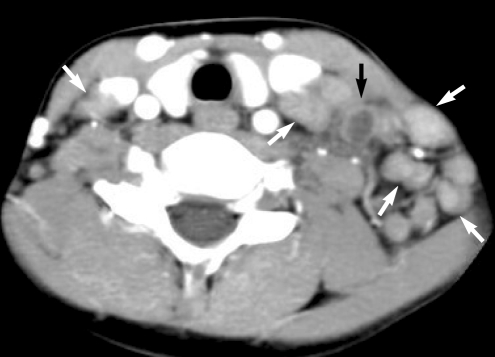
Fig. 3 An 11-yr-old boy with a 2-yr history of acute myeloid leukemia presented with cervical swelling. Post-contrast axial CT image shows multiple homogeneously-enhancing lymph nodes (white arrows) and lymph node with heterogeneous enhancement due to necrosis (black arrow) along the lateral cervical lymph node chains. The lymph node was shown to be a granulocytic sarcoma by local lymph node biopsy.
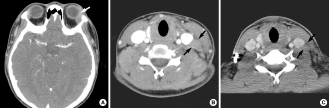
Fig. 4 A 16-yr-old man presented with a left neck mass as an initial manifestation of leukemia. (A) Post-contrast CT scan shows slightly enhancing mass in the superolateral peripheral space of the left orbit. (B) Post-contrast CT scan of the neck shows enlarged and enhancing lymph nodes (arrows) in the lower neck. (C) The lymph nodes are decreased in size on the CT image 7 days after induction chemotherapy.
Cited by 3 articles
-
Clinical characteristics and treatment outcomes of isolated myeloid sarcoma without bone marrow involvement: a single-institution experience
Jung Yeon Lee, Haerim Chung, Hyunsoo Cho, Ji Eun Jang, Yundeok Kim, Soo-Jeong Kim, Jin Seok Kim, Shin Young Hyun, Yoo Hong Min, June-Won Cheong
Blood Res. 2017;52(3):184-192. doi: 10.5045/br.2017.52.3.184.A Case of Myeloid Sarcoma in the Nasal Cavity Occurred in the Patient with Leukemic Transformation in Myelodysplastic Syndrome
Dong Hoo Lee, Sung Yool Park, Ha Young Park, Seong Kook Park
Korean J Otorhinolaryngol-Head Neck Surg. 2020;63(2):81-84. doi: 10.3342/kjorl-hns.2019.00017.Temporal Bone Myeloid Sarcoma
Ki-Hong Chang, Dong-Kee Kim, Beom-Cho Jun, Yong-Soo Park
Clin Exp Otorhinolaryngol. 2009;2(4):198-202. doi: 10.3342/ceo.2009.2.4.198.
Reference
-
1. Guermazi A, Feger C, Rousselot P, Merad M, Benchaib N, Bourrier P, et al. Granulocytic sarcoma (chloroma): imaging findings in adults and children. AJR Am J Roentgenol. 2002; 2. 178(2):319–325. PMID: 11804886.2. Liu PI, Ishimaru T, McGregor DH, Okada H, Steer A. Autopsy study of granulocytic sarcoma (chloroma) in patients with myelogenous leukemia, Hiroshima-Nagasaki 1949-1969. Cancer. 1973; 4. 31(4):948–955. PMID: 4513297.
Article3. Rappaport H. Atlas of tumor pathology, section 3, fascicle 8: tumors of the hematopoeitic system. 1966. Washington (DC): Armed Forces Institute of Pathology.4. Ooi GC, Chim CS, Khong PL, Au WY, Lie AK, Tsang KW, et al. Radiologic manifestations of granulocytic sarcoma in adult leukemia. AJR Am J Roentgenol. 2001; 6. 176(6):1427–1431. PMID: 11373207.
Article5. Pui MH, Fletcher BD, Langston JW. Granulocytic sarcoma in childhood leukemia: imaging features. Radiology. 1994; 3. 190(3):698–702. PMID: 8115614.
Article6. Lee YH, Lee NJ, Choi EJ, Kim JH. Granulocytic sarcoma (chloroma) presenting as a lateral neck mass: initial manifestation of leukemia: a case report. Eur Arch Otorhinolaryngol. 2006; 1. 263(1):16–18. PMID: 16205903.
Article7. Lee B, Fatterpekar GM, Kim W, Som PM. Granulocytic sarcoma of the temporal bone. AJNR Am J Neuroradiol. 2002; 10. 23(9):1497–1499. PMID: 12372738.8. Nayak DR, Balakrishnan R, Raj G, Pillai S, Rao L, Manohar C. Granulocytic sarcoma of the head and neck: a case report. Am J Otolaryngol. 2001; Jan–Feb. 22(1):80–83. PMID: 11172221.
Article9. Som PM, Brandwein MS. Som PM, Curtin DH, editors. Lymph Nodes. Head and neck imaging. 2003. 4th ed. St. Louis (MO): Mosby;p. 1805–2003.10. Mukherji SK. Som PM, Curtin DH, editors. Pharynx. Head and neck imaging. 2003. 4th ed. St. Louis (MO): Mosby;p. 1465–1520.
- Full Text Links
-
- Actions
-
Cited
- CITED
-
- Close
- Share
-
- Similar articles
-
- A Case of Granulocytic Sarcoma Presenting as a Head and Neck Neoplasm
- Granulocytic Sarcoma in the Leg Mimicking Hemorrhagic Abscess
- A case report:the granulocytic sarcoma in the head and neck
- Undifferentiated Embryonal Sarcoma of Liver in Child
- A Case of Isolated Granulocytic Sarcoma of the Ovary in Nonleukemic Patients
