A Case Report of Secretan's Disease in Both Hands
- Affiliations
-
- 1Department of Radiology, Kyungpook National University Hospital, Kyungpook National University School of Medicine, Daegu, Korea. yijh@knu.ac.kr
- KMID: 2002904
- DOI: http://doi.org/10.3348/jksr.2013.68.6.511
Abstract
- Secretan's disease is a rare clinical condition with recurrent or persistent hard swelling and hyperplasia on the dorsal aspect of the hand with peritendinous fibrosis around the extensor tendons. Although it is usually believed to involve a hand with preceding traumatic injury, here we present a case of Secretan's disease occurring in both hands without obvious trauma history in a 73-year-old female, with clinical, radiological, and histological findings. The magnetic resonance imaging exhibited an ill-defined infiltrative mass-like lesion with homogenous dark signal intensity on T1 and T2-weighted images in the dorsal subcutaneous layer of the hand. The lesion showed no obvious enhancement during contrast-enhanced T1-weighted images. Microscopic examination of surgical specimen indicated marked fibrosis.
Figure
-
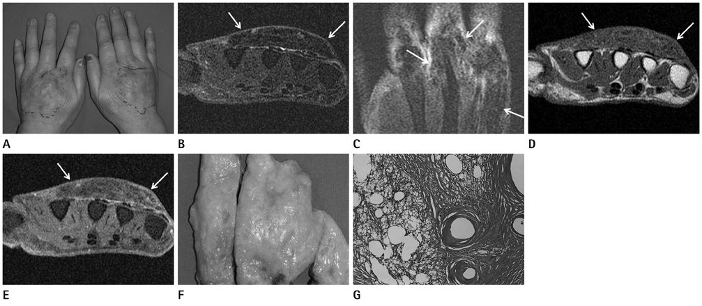
Fig. 1 A 73-year-old woman diagnosed with Secretan's disease. A. Clinical photograph shows localized swelling with mild erythematous skin change in the dorsal metacarpal area of both hands. B, C. Axial (B) and coronal (C) fat-suppressed T2-weighted images of left hand show the diffuse hypointense infiltrating mass-like lesion (arrows) in the subcutaneous layer on the dorsal metacarpal area and peritendinous area around the metacarpophalangeal joints. D, E. The lesion shows also diffuse low signal intensity on T1-weighted image (D) and no vivid enhancement on axial (E) contrast-enhanced images (arrows) of the left hand. F. Photograph of the surgical specimen shows a yellowish, firm soft tissue lesion with lobulated irregular margin. G. Photomicrograph (hematoxylin-eosin, × 100) shows dense fibrosis with prominent fatty degeneration and no obvious evidence of a neoplasm.
Reference
-
1. Moretta DN, Cooley RD Jr. Secrétan's disease: a unique case report and literature review. Am J Orthop (Belle Mead NJ). 2002; 31:524–527.2. Whitney TM, Jones NF. Magnetic resonance imaging findings in Secretan's disease. J Hand Surg Am. 1995; 20:464–466.3. Reading G. Secretan's syndrome: hard edema of the dorsum of the hand. Plast Reconstr Surg. 1980; 65:182–187.4. Saferin EH, Posch JL. Secretan's disease: post-traumatic hard edema of the dorsum of the hand. Plast Reconstr Surg. 1976; 58:703–707.5. Ramos JA, Bush D, Harrington TM. Secretan's syndrome. Orthopedics. 2007; 30:239–240.6. Angelini G, Meneghini CL, Vena GA. Secretan's syndrome: an artefact oedema of the hand. Contact Dermatitis. 1982; 8:345–346.7. Abnousi F, Chou LB. Secretan's disease of the foot: a case report and review. Foot Ankle Int. 2008; 29:248–250.
