Breast Metastasis from Malignant Paraganglioma: A Case Report
- Affiliations
-
- 1Department of Radiology and Center for Imaging Science, Samsung Medical Center, Sungkyunkwan University School of Medicine, Seoul, Korea. bkhan@skku.edu
- 2Department of Radiology, Gil Hospital, Gachon University of Medicine and Science, Incheon, Korea.
- KMID: 2002902
- DOI: http://doi.org/10.3348/jksr.2013.68.6.499
Abstract
- We report a case of metastatic breast cancer from paraganglioma and describe the radiologic findings in a 32-year-old woman who had a history of excision for carotid body paraganglioma. Breast metastasis from malignant paraganglioma showed a well-defined mass with hypoechogenicity, posterior acoustic enhancement and increased vascularity on ultrasonography, and strong enhancements on contrast-enhanced MRI and diffusion restriction on diffusion weighted image.
MeSH Terms
Figure
-
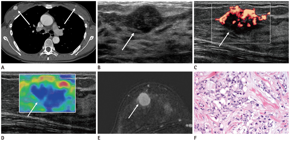
Fig. 1 A 32-year-old woman with metastatic paraganglioma of the right breast. A. Axial contrast-enhanced chest CT scan shows two enhancing nodules in both breasts (arrows). B. Ultrasonography (US) shows a 1.8 cm sized, oval shape, circumscribed, hypoechoic mass with posterior acoustic enhancement in the right upper center (arrow). C. The mass shows high vascularity on power Doppler US (arrow). D. Elastographic image shows predominantly blue with an elasticity score of 4 (arrow). E. Axial dynamic contrast-enhanced fat-suppressed T1-weighted image MRI shows strong enhancement of the mass (arrow). F. Photomicrograph shows typical pattern of cell nests separated by prominent fibrous bands. Nests of chief cells in tumor are separated by fibrous bands (haematoxylin and eosin stained; original magnification, × 400).
Reference
-
1. Sandison AT. Metastatic tumours in the breast. Br J Surg. 1959; 47:54–58.2. Hajdu SI, Urban JA. Cancers metastatic to the breast. Cancer. 1972; 29:1691–1696.3. Paulus DD, Libshitz HI. Metastasis to the breast. Radiol Clin North Am. 1982; 20:561–568.4. Toombs BD, Kalisher L. Metastatic disease to the breast: clinical, pathologic, and radiographic features. AJR Am J Roentgenol. 1977; 129:673–676.5. Vergier B, Trojani M, de Mascarel I, Coindre JM, Le Treut A. Metastases to the breast: differential diagnosis from primary breast carcinoma. J Surg Oncol. 1991; 48:112–116.6. Vaughan A, Dietz JR, Moley JF, Debenedetti MK, Aft RL, Gillanders WE, et al. Metastatic disease to the breast: the Washington University experience. World J Surg Oncol. 2007; 5:74.7. Williams SA, Ehlers RA 2nd, Hunt KK, Yi M, Kuerer HM, Singletary SE, et al. Metastases to the breast from nonbreast solid neoplasms: presentation and determinants of survival. Cancer. 2007; 110:731–737.8. Lee JH, Kim SH, Kang BJ, Cha ES, Kim HS, Choi JJ. Metastases to the breast from extramammary malignancies-sonographic features. J Clin Ultrasound. 2011; 39:248–255.9. Kliewer KE, Wen DR, Cancilla PA, Cochran AJ. Paragangliomas: assessment of prognosis by histologic, immunohistochemical, and ultrastructural techniques. Hum Pathol. 1989; 20:29–39.10. Lee JH, Barich F, Karnell LH, Robinson RA, Zhen WK, Gantz BJ, et al. National Cancer Data Base report on malignant paragangliomas of the head and neck. Cancer. 2002; 94:730–737.11. Rao AB, Koeller KK, Adair CF. Armed Forces Institute of Pathology. From the archives of the AFIP. Paragangliomas of the head and neck: radiologic-pathologic correlation. Radiographics. 1999; 19:1605–1632.
- Full Text Links
-
- Actions
-
Cited
- CITED
-
- Close
- Share
-
- Similar articles
-
- A case report of malignant paraganglioma with hepatic metastases
- A Case Report of Recurrent Malignant Paraganglioma with Lung and Spine Metastasis that Occurred Sixteen Years after Primary Tumor Excision
- A Case of Metastatic Malignant Paraganglioma Causing Spinal Root Compression
- Malignant Paraganglioma in the Common Hepatic Duct
- Pancreatic metastasis from malignant phyllodes tumor of the breast
