Aneurysm of the Common Iliac Vein Mimicking a Pelvic Mass
- Affiliations
-
- 1Department of Radiology, Chosun University College of Medicine, Gwangju, Korea. dhk0827@chosun.ac.kr
- 2Department of Anesthesiology and Pain Medicine, Seonam University College of Medicine, Namwon, Korea.
- KMID: 2002888
- DOI: http://doi.org/10.3348/jksr.2013.69.1.39
Abstract
- Venous aneurysm, especially of primary origin, is rare. The authors report a case of a 63-year-old female who was admitted for back pain and an aneurysm of the common iliac which was detected incidentally. CT, magnetic resonance (MR), Doppler ultrasonography, and conventional venography showed an aneurysm of the left common iliac vein measuring 4.5 x 3 x 4 cm. Because there were no complications of the aneurysm, no further treatment was administered. Herein, we describe findings of a venous aneurysm of the common iliac vein mimicking a pelvic mass on CT and MR scans and with a review of the literature.
MeSH Terms
Figure
-
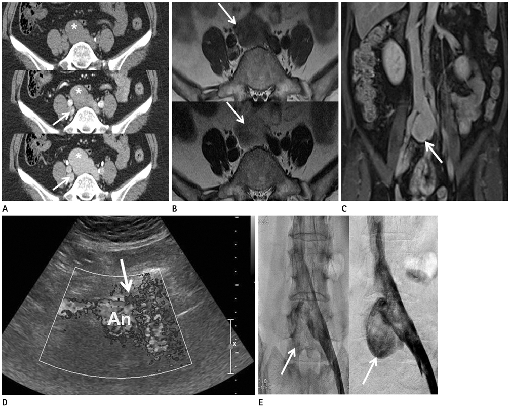
Fig. 1 A 63-year-old female with a venous aneurysm of the common iliac vein. A. Abdominopelvic CT scans (upper image in non-enhanced scan, middle and lower images in portal and delayed phases, respectively) show heterogeneous enhancement of the tumor (asterisks) relative to the common iliac vein (arrows). B. T1-weighted axial scan (upper image) shows a hypointense mass (arrows) relative to muscle and common iliac vein. T2-weighted scan (lower image) shows a hyperintense mass with central hypointensity. C. T1-weighted coronal scan with contrast enhancement shows the same enhancement of the tumor (arrow) compared with the common iliac vein. D. Sonography with color doppler shows blood inflow to the aneurysm (An) and aneurysmal neck (arrow). E. Early (right image) and late (left image) phases of venogram show contrast filling in the aneurysm (arrows) on the anteromedial aspect of the left.
Reference
-
1. Gillespie DL, Villavicencio JL, Gallagher C, Chang A, Hamelink JK, Fiala LA, et al. Presentation and management of venous aneurysms. J Vasc Surg. 1997; 26:845–852.2. Ysa A, Bustabad MR, Arruabarrena A, Pérez E, Gainza E, Alonso JA. Thrombosed iliac venous aneurysm: a rare form of presentation of a congenital anomaly of the inferior vena cava. J Vasc Surg. 2008; 48:218–222.3. Abbott OA, Leigh TF. Aneurysmal dilatations of the superior vena caval system. Ann Surg. 1964; 159:858–872.4. Banno H, Yamanouchi D, Fujita H, Nagata J, Kobayashi M, Matsushita M, et al. External iliac venous aneurysm in a pregnant woman: a case report. J Vasc Surg. 2004; 40:174–178.5. Irace L, Gossetti B, Benedetti-Valentini F, Francalanci P, Gallo P. Aneurysm of the left renal vein: a case report. J Vasc Surg. 1994; 19:943–944.6. Sam AD 2nd, Frusha JD, McNeil JW, Olinde AJ. Repair of a blunt traumatic inferior vena cava laceration with commercially available endografts. J Vasc Surg. 2006; 43:841–843.7. Rajiah P, Sinha R, Cuevas C, Dubinsky TJ, Bush WH Jr, Kolokythas O. Imaging of uncommon retroperitoneal masses. Radiographics. 2011; 31:949–976.8. Krinsky G, Johnson G, Rofsky N, Shapiro R, Yang B, Weinreb J. Venous aneurysms: MR diagnosis with the "layered gadolinium" sign. J Comput Assist Tomogr. 1997; 21:623–627.
- Full Text Links
-
- Actions
-
Cited
- CITED
-
- Close
- Share
-
- Similar articles
-
- A Case of Iliac Artery Aneurysm Masquerading as Pelvic Sarcoma
- Saccular Aneurysm of Intraheparic Portal Vein Mimicking Hypervascular Hepatic Mass: A Case Report
- Massive retroperitoneal hemorrhage with a false aneurysm of left common iliac artery complicating lumbardiskectomy
- Spontaneous Rupture of Aortoiliac Aneurysm into Left Common Iliac Vein Combined with Gastric Leiomyoma: A case report
- Surgical anatomy of the common iliac veins during para-aortic and pelvic lymphadenectomy for gynecologic cancer
