CT and Magnetic Resonance Imaging Findings of Lipomatous Hemangiopericytoma of Skull Base: A Case Report
- Affiliations
-
- 1Department of Radiology, Eulji University Hospital, Daejeon, Korea. midosyu@eulji.ac.kr
- 2Department of Neurosurgery, Eulji University Hospital, Daejeon, Korea.
- 3Department of Pathology, Eulji University Hospital, Daejeon, Korea.
- KMID: 2002883
- DOI: http://doi.org/10.3348/jksr.2013.69.1.1
Abstract
- Lipomatous hemangiopericytoma (LHPC) is recently recognized as a rare hemangiopericytoma variant. To our knowledge, imaging features of LHPC involving skull base have not yet been reported. We present the imaging features of LHPC of skull base in a 44-year-old female, along with a literature review CT and magnetic resonance imagings showed well-enhanced fatty issues containing temporal skull base masses, with pressure bony erosions.
MeSH Terms
Figure
-
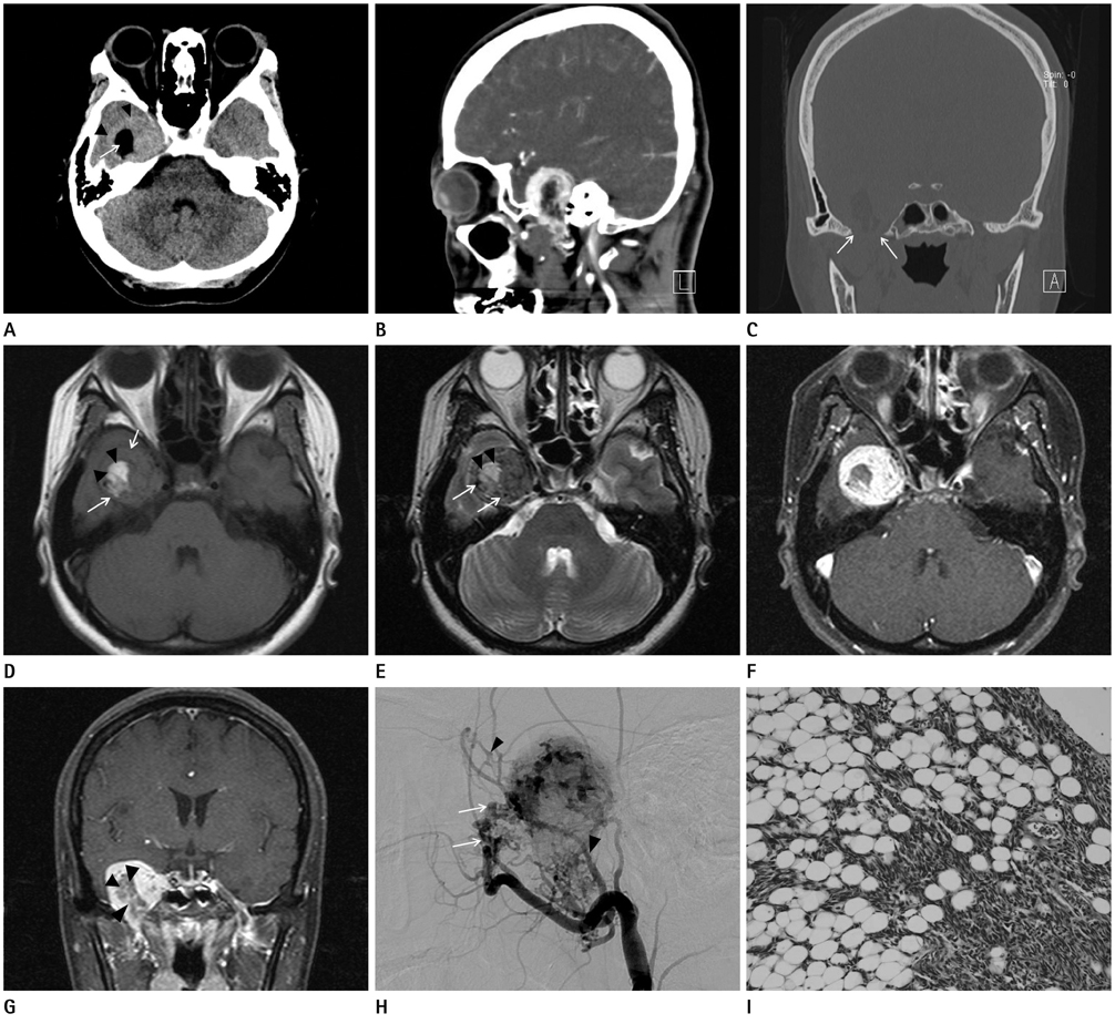
Fig. 1 A 44-year-old woman who presented with 1 month history of dizziness. A. Precontrast CT image shows well-defined round solid increased attenuated tumor (arrowheads), with containing intratumoral fat-attenuated (-60 Hounsfield unit) component (arrow) at the right temporal base area. B. Sagittal postcontrast CT image shows well enhancing solid hypervascular extraaxial tumor with small inferior extracranial extention via widened foramen ovale. C. Coronal temporal bone CT image shows tumor with adjacent bone pressure erosion and small inferior extracranial extention via widened foramen ovale (arrows). D. T1-weighted axial magnetic resonance (MR) image shows slightly hypointense mass (arrows) with central irregular high signal intensity portions, intratumoral lipoma component (arrowheads). E. T2-weighted axial MR image shows intermediate signal intensity mass, intratumoral signal void structures (arrows), with intratumoral high signal intensity portions (arrowheads). F. Contrast-enhanced T1-weighted axial MR image shows well enhancing solid mass. G. Contrast-enhanced T1-weighted coronal MR image shows small inferior extension to extracranial space via foramen ovale with central fat-saturated poorly enhancing portion (arrowheads). H. Lateral angiography shows hypervascular tumor staining fed by the right middle meningeal artery (arrowheads), and internal maxillary artery (arrows). I. Histologic features of lipomatous hemangiopericytoma. Microscopic view shows a highly vascular tumor, with hyperchromatic nuclei and normal adipocytes are seen within the tumor matrix (H&E, × 200).
Reference
-
1. Chiechi MV, Smirniotopoulos JG, Mena H. Intracranial hemangiopericytomas: MR and CT features. AJNR Am J Neuroradiol. 1996; 17:1365–1371.2. Fountas KN, Kapsalaki E, Kassam M, Feltes CH, Dimopoulos VG, Robinson JS, et al. Management of intracranial meningeal hemangiopericytomas: outcome and experience. Neurosurg Rev. 2006; 29:145–153.3. Nielsen GP, Dickersin GR, Provenzal JM, Rosenberg AE. Lipomatous hemangiopericytoma. A histologic, ultrastructural and immunohistochemical study of a unique variant of hemangiopericytoma. Am J Surg Pathol. 1995; 19:748–756.4. Folpe AL, Devaney K, Weiss SW. Lipomatous hemangiopericytoma: a rare variant of hemangiopericytoma that may be confused with liposarcoma. Am J Surg Pathol. 1999; 23:1201–1207.5. Shaia WT, Bojrab DI, Babu S, Pieper DR. Lipomatous hemangiopericytoma of the skull base and parapharyngeal space. Otol Neurotol. 2006; 27:560–563.6. Servo A, Jääskeläinen J, Wahlström T, Haltia M. Diagnosis of intracranial haemangiopericytomas with angiography and CT scanning. Neuroradiology. 1985; 27:38–43.7. Chen Q, Chen XZ, Wang JM, Li SW, Jiang T, Dai JP. Intracranial meningeal hemangiopericytomas in children and adolescents: CT and MR imaging findings. AJNR Am J Neuroradiol. 2012; 33:195–199.8. Jing HB, Meng QD, Tai YH. Lipomatous hemangiopericytoma of the stomach: a case report and a review of literature. World J Gastroenterol. 2011; 17:4835–4838.9. Jääskeläinen J, Servo A, Haltia M, Wahlström T, Valtonen S. Intracranial hemangiopericytoma: radiology, surgery, radiotherapy, and outcome in 21 patients. Surg Neurol. 1985; 23:227–236.10. Buetow MP, Buetow PC, Smirniotopoulos JG. Typical, atypical, and misleading features in meningioma. Radiographics. 1991; 11:1087–1106.
