Lipoma-like Liposarcoma with Osteosarcomatous Dedifferentiation of the Chest Wall: A Case Report
- Affiliations
-
- 1Department of Radiology, College of Medicine, Korea University, Korea. mallecot@hanmail.net
- 2Department of Pathology, College of Medicine, Korea University, Korea.
- KMID: 2000054
- DOI: http://doi.org/10.13104/jksmrm.2011.15.3.251
Abstract
- We report a case of liposarcoma with osteosarcomatous dedifferentiation of the chest wall in a 58-year-old man, which had been initially mistaken as myositis ossificans. CT and MRI demonstrated a soft tissue mass consisting of two components: a non-lipomatous area with amorphous calcification/ossification and a well-encapsulated fatty component. Based on local excision in the non-lipomatous area, myositis ossificans was initially diagnosed. As the mass was gradually enlarging, however, wide excision including the fatty component was performed and histological assessment revealed lipoma-like, well-differentiated liposarcoma with high-grade osteosarcomatous dedifferentiation. Here, we describe the radiological-pathological features of this rare neoplasm.
Keyword
Figure
-
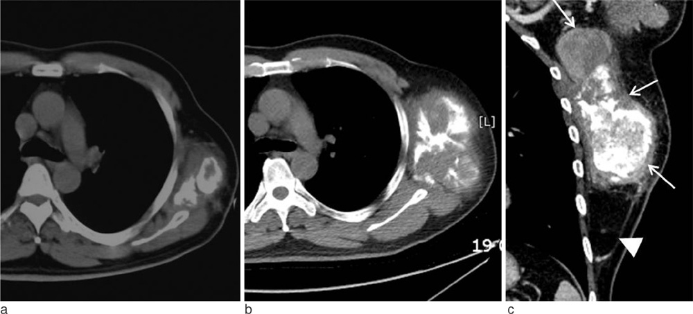
Fig. 1 Lipoma-like liposarcoma with osteosarcomatous dedifferentiation of the chest wall in a 58-year-old man. Non-enhanced chest CT scan (a) demonstrates a soft tissue mass with discrete central ossification/calcification at his lateral chest wall. On follow-up CT scan taken after 9 months from (a), an axial image (b) demonstrates significant increase in the tumor size. Coronal reformatted imaging (c) shows an elongated bimorphic mass with a length of 18 cm. The larger upper portion (arrows) of the mass shows non-lipomatous soft tissue attenuation containing multiple amorphous areas of calcification/ossification. The fatty lower portion (arrowhead) shows no definite solid component.

Fig. 2 Lipoma-like liposarcoma with osteosarcomatous dedifferentiation of the chest wall in a 58-year-old man. Coronal T1-weighted image (a) of the chest reveals that the lower portion of the tumor is of homogenous high signal intensity (arrowhead), consistent with fat, whereas the larger upper portion (asterisk) shows heterogeneous intermediate and low signal intensity with subtle high signal intensity of fat (arrows) in the periphery. Corresponding STIR image (b) and fat-saturated coronal T1-weighted image (c) after intravenous gadolinium administration confirm the fatty nature of the lower portion (arrowheads) as well as the periphery (arrows) of the upper portion of the tumor by nulling the signal return. There is no enhancement in the fatty lower portion of the tumor other than thin capsular enhancement. The upper portion of the tumor (asterisk) shows heterogeneous enhancement and heterogeneous T2 signal from bright high signal to dark low signal representing cystic changes and tumor mineralization, respectively.

Fig. 3 (a) Photomicrograph (original magnification, ×100; hematoxylin-eosin stain) of the lipomatous portion of the tumor shows predominantly lipocytes admixed with scattered atypical stromal cells (arrows) with irregular, hyperchromatic nuclei. (b) Photomicrograph (original magnification, ×40; hematoxylin-eosin stain) of the osteosarcomatous component demonstrates dense trabecular bony spicules. (c) In a magnified view of (b) (×200), there are malignant spindle cells with hyperchromatic nuclei (arrows) with osteoid and woven bone formation.
Reference
-
1. Ippolito V, Brien EW, Menendez LR, Mirra JM. Case report 797: "Dedifferentiated" lipoma-like liposarcoma of soft tissue with focal transformation to high-grade "sclerosing" osteosarcoma. Skeletal Radiol. 1993. 22:604–608.2. Evans HL, Khurana KK, Kemp BL, Ayala AG. Heterologous elements in the dedifferenatiated component of dedifferentiated liposarcoma. Am J Surg Pathol. 1994. 18:1150–1157.3. Henricks WH, Chu YC, Goldblum JR, Weiss SW. Dedifferentiated liposarcoma: a clinicopathological analysis of 155 cases with a proposal for an expanded definition of dedifferentiation. Am J Surg Pathol. 1997. 21:271–281.4. Yamamoto T, Matsushita T, Marui T, et al. Dedifferenatiated liposarcoma with chondrobalstic osteosarcomatous dedifferenatiation. Pathology International. 2000. 50:558–561.5. Takanami I, Imamura T. Dedifferentiated liposarcoma of the pleura: report of a case. Surg Today. 2005. 313–316.6. Toms A, White LM, Kandel R, Bell R. Low-grade liposarcoma with osteosarcomatous dedifferentiation: radiological and histological features. Skeletal Radiol. 2003. 32:286–289.7. Yu L, Fung S, Hojnowski L, Damron T. Dedifferentiated liposarcoma of soft tissue with high-grade osteosarcomatous dedifferentiation. Radiographics. 2005. 25:1082–1086.8. Toshiyasu T, Ehara S, Yamaguchi T, Nishida J, Shiraishi H. Dedifferentiated liposarcoma of the retroperitoneum with osteosarcomatous component: report of two cases. Clinical Imaging. 2009. 33:70–74.9. Yoshida A, Ushiku T, Motoi T, Tatsuhiro T, Fukayama M, Tsuda H. Well-differentiated liposarcoma with low-grade osteosarcomatous component. Am J Surg Pathol. 2010. 34(9):1361–1366.10. Evans HL. Liposarcoma: a study of 55 cases with reassessment of its classification. Am J Surg Pathol. 1979. 3:507–523.
- Full Text Links
-
- Actions
-
Cited
- CITED
-
- Close
- Share
-
- Similar articles
-
- Erratum: Lipoma-like Liposarcoma with Osteosarcomatous Dedifferentiation of the Chest Wall: A Case Report
- Magnetic Resonance Imaging Features That Permit Differential Diagnosis of Chest Wall Liposarcoma Mimicking Lipoma in Men
- Unusual bilateral vulvar liposarcoma
- A Case of Myxoid Liposarcoma of Neck
- A Case of Retroperitoneal Liposarcoma
