J Korean Acad Conserv Dent.
2002 Jul;27(4):389-393. 10.5395/JKACD.2002.27.4.389.
The effect of the endodontic access cavity on the marginal leakage of crowns
- Affiliations
-
- 1Department of Conservative Dentistry, Yonsei University, Korea.
- 2Department of Restorative Dentistry, University of Pennsylvania, USA.
- KMID: 1987294
- DOI: http://doi.org/10.5395/JKACD.2002.27.4.389
Abstract
- The marginal integrity of the crown can be broken during endodontic access cavity preparation due to the vibration of burs. Therefore, the purpose of this study was to evaluate the effect of endodontic access cavity preparation on the marginal leakage of full veneer gold crowns. 24 intact molars were mounted in acrylic resin blocks and prepared for crowns by a restorative dentist and crowns were cast with gold alloy. 20 Crowns were cemented with glass ionomer cement and 2 crowns were not cemented for positive control. 200 thermo-cycles from 5degrees C to 50degrees C with a travel time of 20s were completed. Then samples were randomly divided into 2 experimental groups of 9 each. Endodontic access preparation and zinc-oxide eugenol temporary fillings were done in Group 1. Teeth in Group 2 were not treated. Samples were coated with 2 layers of nail varnish and were immersed in 1% methylene blue dye for 20 hrs. Endodontic access was prepared in 2 samples, which were coated with nail varnish on all surfaces for negative control. After washing in running water, gold crowns were cut with a #330 bur. Four buccolingual sections, 2 mm apart, were cut from the central section of each tooth and were examined and scored under the microscope for dye leakage. Score 1: leakage to the cervical 1/3 of the axial wall, Score 2: leakage to the middle 1/3 of the axial wall, Score 3: leakage to the coronal 1/3 of the axial wall, Score 4: leakage to the occlusal surface. The median value for Group 1 is 4 and for Group 2 is 2. The result of this study showed that samples in Group 1 leaked more than those in Group 2. This finding was significant(P<0.001).
Keyword
MeSH Terms
Figure
-

Fig. 1 Schematic drawing of section for leakage observation.
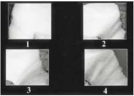
Fig. 2 Scoring criteria of dye leakage(×25).
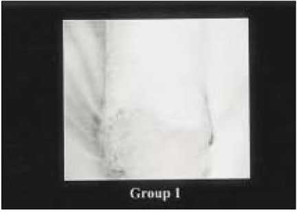
Fig. 3 Leakage of group I sample scoring 4 which is leakage to the occlusal surface(×25).
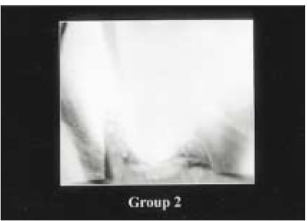
Fig. 4 Leakage of group II sample scoring 2 which is leakage to the middle 1/3 of the axial wall(×25).
Reference
-
1. Saunders WP, Saunders EM. Coronal leakage as a cause of failure in root canal therapy: a review. Endod Dent Traumatol. 1994. 10:105–108.2. Ingle JI, Taintor JF. Endodontics. 1985. 3rd ed. Philadelphia: Lea & Febiger;27–50.3. Swartz DB, Skidmore AE, Griffin JA. Twenty years of endodontic success and failure. J Endod. 1983. 9:198–202.
Article4. Vire DE. Failure of endodontically treated teeth: classification and evaluation. J Endod. 1991. 17:338–342.
Article5. Ray HA, Trope M. Periapical status of endodontically treated teeth in relation to the technical quality of the root filling and the coronal restoration. Int Endod J. 1995. 28:12–18.
Article6. Goldman M, Laosonthorn P, White RR. Microleakage-full crown and the dental pulp. J Endod. 1992. 18:473–475.7. Madison S, Jordan RD, Krell KV. The effects of rubber dam retainers on porcelain fused-to-metal restorarions. J Endod. 1986. 12:183–186.
Article8. Yu YC, Abbott PV. The effect of endodontic access cavity preparation and subsequent restorative procedures on incisor crown retention. Aust Dent J. 1994. 39:247–251.
Article9. Kakehashi S, Stanley HR, Fitzgerald RJ. The effect of surgical exposures of dental pulps in germ-free and conventional laboratory rats. Oral Surg. 1965. 20:340–349.
Article10. Sundqvist G. Bacteriological studies of necrotic dental pulps. 1976. (No.7):1–94. Thesis. Umea Univ.Odont.Diss.11. Möller AJ, et al. Influence on periapical tissues of indigenous oral bacteria and necrotic pulp tissue in monkeys. Scand J Dent Res. 1981. 89:475–484.
Article12. Kim S, et al. Functional alterations in pulpal microcirculation in response to various dental procedures and materials. Proc Finn Dent Soc. 1992. 88:65–71.13. Langeland K, Langeland LK. Pulp reactions to crown preparation, impression, temporary crown fixation and permanent cementation. J Prosthet Dent. 1965. 15:129–143.
Article14. Felton D, et al. Long term effect of crown preparation on pulp viability. J Dent Res. 1989. 68:1009.15. Sundqvist G, et al. Microbiologic analysis of teeth with failed endodontic treatment and the outcome of conservative re-treatment. Oral Surg Oral Med Oral Pathol Oral Radiol Endod. 1998. 85:86–93.
Article16. Hsu YY, Kim S. The resected root surface: The issue of canal isthmuses. Dent Clin North Am. 1997. 41:529–540.17. Kersten HW, Moorer WR. Particles and molecules in endodontic leakage. Int Endod J. 1989. 22:118–124.
Article18. Goldman M, Simmonds S, Rush R. The usefulness of dye penetration studies re-examined. Oral Surg. 1989. 67:327–332.19. Starkey DL, Anderson RW, Pashley DH. An evaluation of the effect of methylene blue dye pH on apical leakage. J Endod. 1993. 19:435–439.
Article
- Full Text Links
-
- Actions
-
Cited
- CITED
-
- Close
- Share
-
- Similar articles
-
- Fracture resistance and marginal fit of the zirconia crowns with varied occlusal thickness
- Traditional and minimally invasive access cavities in endodontics: a literature review
- Marginal accuracy and fracture strength of Targis/Vectris Crowns prepared with different preparation designs
- Marginal fit of the digident CAD/CAM zirconia ceramic crowns
- The influence of different access cavity designs on the fracture strength in endodontically treated mandibular anterior teeth
