The effect of smear layer treatment on the microleakage
- Affiliations
-
- 1Department of Conservative Dentistry, Division of Dentistry, Graduate school of Kyung Hee University, Seoul, Korea. gwchoi@khu.ac.kr
- KMID: 1986881
- DOI: http://doi.org/10.5395/JKACD.2006.31.5.378
Abstract
- The purpose of this study was to compare the sealing ability of root canal obturation with or without the treatment of smear layer. Eighty extracted human teeth with one canal were selected. Instrumentation was performed with crown-down technique. After instrumentation, root canals of the NaOCl group and NaOCl-6 group were irrigated with 3% NaOCl. EDTA group and EDTA-6 group were irrigated with 17% EDTA. Then all teeth were obturated using continuous wave obturation technique. NaOCl group and EDTA group were immersed in methylene blue solution for 84hours. NaOCl-6 group and EDTA-6 group were immersed in methylene blue solution for 6months. The teeth were sectioned at 1.5 mm (Level 1), 3.0 mm (Level 2) and 4.5 mm (Level 3) from the root apex. The length of dye-penetrated interface and the circumferential length of canal at each level were measured using Sigma-Scan Pro 5.0. 1. The mean leakage ratio was decreased cervically. 2. NaOCl group showed higher mean leakage ratio than EDTA group at each level. But there was significant difference at level 1 only (p < 0.05). 3. NaOCl-6 group showed higher mean leakage ratio than EDTA-6 group at each level. But there was significant difference at level 1 only (p < 0.05). 4. NaOCl-6 group showed higher mean leakage ratio than NaOCl group at each level. But there was significant difference at level 1 only (p < 0.05). 5. EDTA-6 group showed higher mean leakage ratio than EDTA group at each level. But there was no significant difference. 6. In NaOCl group and NaOCl-6 group, scanning electron micrographs of tooth sections generally covered with smear layer. In EDTA group and EDTA-6 group, tooth sections showing the penetration of sealers to opened dentinal tubules. The results suggest that removal of smear layer was effective to reduce the apical microleakage of the root canal.
Keyword
MeSH Terms
Figure
-
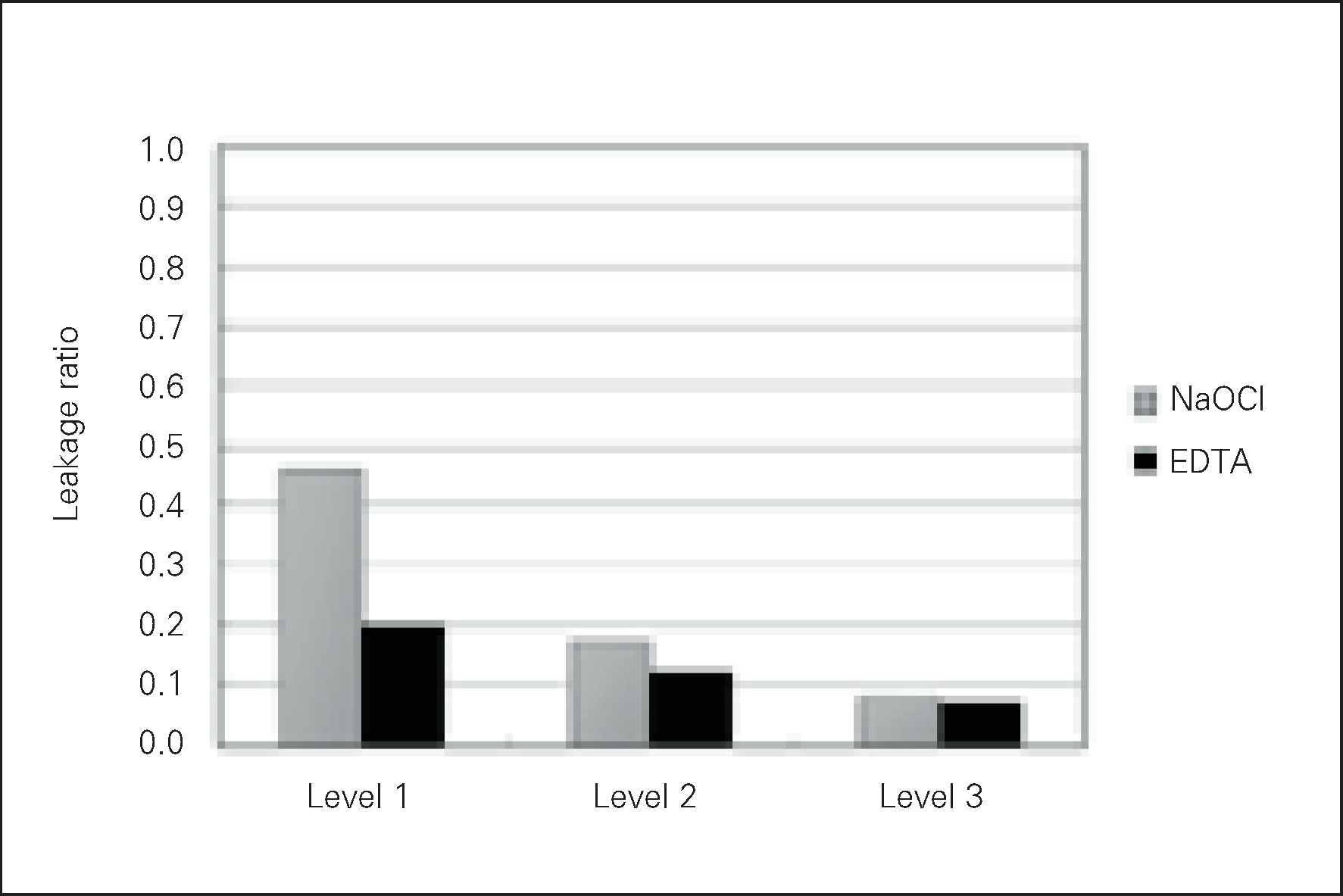
Figure 1. Mean leakage ratio in NaOCl group and EDTA group.

Figure 2. Mean leakage ratio in NaOCl-6 group and EDTA-6 group.

Figure 3. Mean leakage ratio in NaOCl group and NaOCl-6 group.

Figure 4. Mean leakage ratio in EDTA group and EDTA-6 group.

Figure 5. Photograph of the NaOCl group Level 1 (× 120).

Figure 6. Photograph of the NaOCl group Level 2 (× 120).

Figure 7. Photograph of the NaOCl group Level 3 (× 120).

Figure 8. Photograph of the EDTA group Level 1 (× 120).
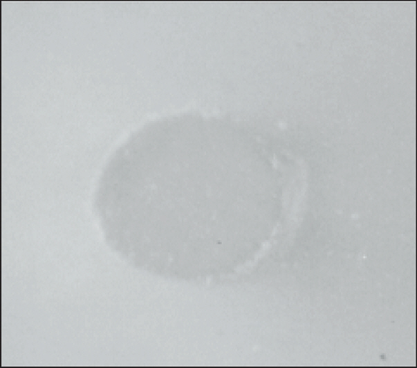
Figure 9. Photograph of the EDTA group Level 2 (× 120).

Figure 10. Photograph of the EDTA group Level 3 (× 120).
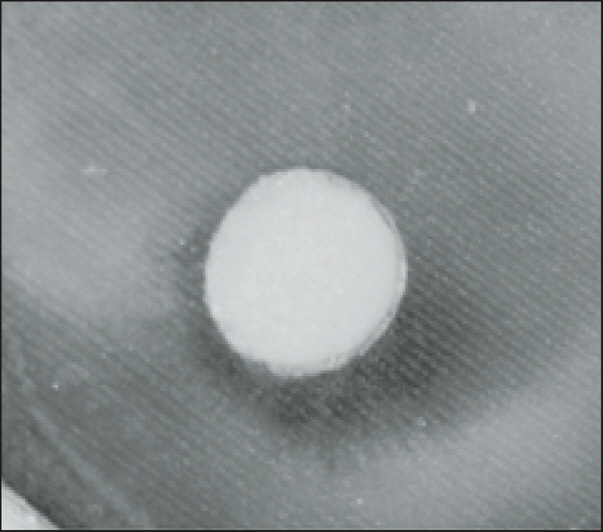
Figure 11. Photograph of the NaOCl-6 group Level 1 (× 120).

Figure 12. Photograph of the NaOCl-6 group Level 2 (× 120).
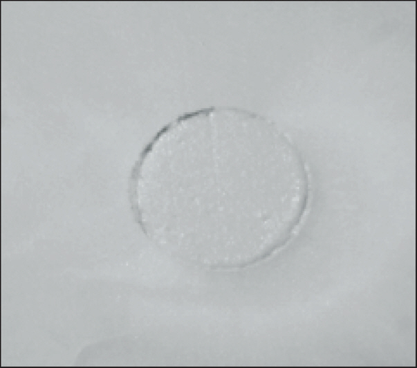
Figure 13. Photograph of the NaOCl-6 group Level 3 (× 120).
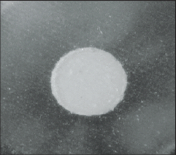
Figure 14. Photograph of the EDTA-6 group Level 1 (× 120).
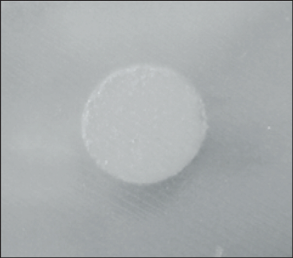
Figure 15. Photograph of the EDTA-6 group Level 2 (× 120).
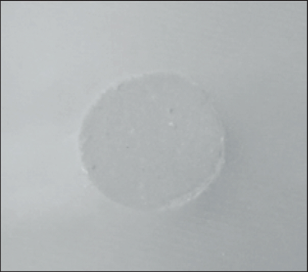
Figure 16. Photograph of the EDTA-6 group Level 3 (× 120).
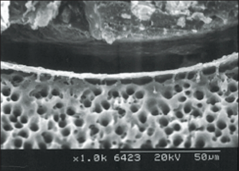
Figure 17. Photograph of the NaOCl group Level 1 (× 1000); Scanning electron micrographs of tooth sections showing the gaps between gutta-percha and the surface of root canals. Surface generally covered with smear layer. Gaps were occuring between the smear layer and the gutta-percha.
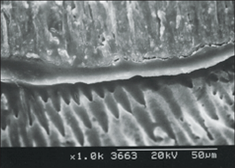
Figure 18. Photograph of the NaOCl group Level 2 (× 1000); Scanning electron micrographs of tooth sections showing the gap-free region between gutta-percha and the surface of root canals. Surface generally covered with smear layer.

Figure 19. Photograph of the EDTA group Level 1 (× 1000); Scanning electron micrographs of tooth sections showing the penetration of sealers to opened dentinal tubules. Gap-free region was showing between the sealer and the dentinal tubules. Numerous sealer tags could be seen within the intertubular dentin. But, voids were occuring between the sealer and the gutta-percha.
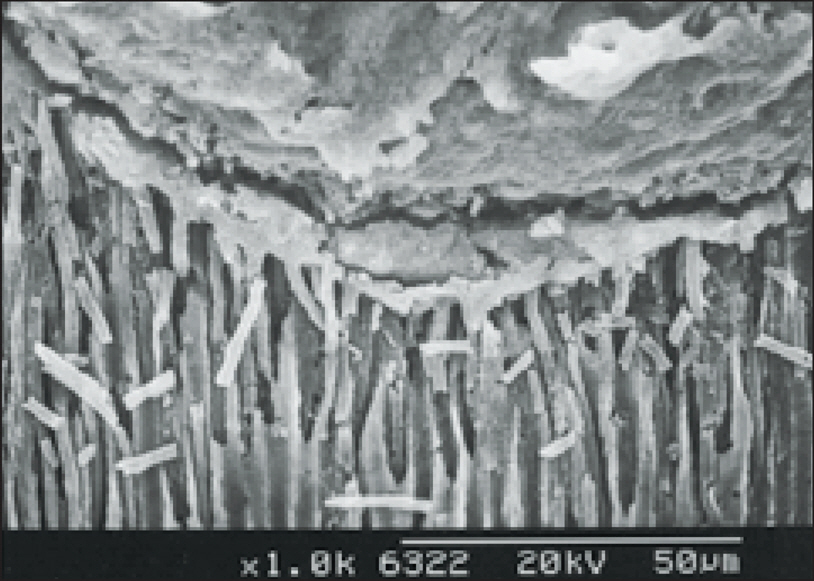
Figure 20. Photograph of the EDTA group Level 2 (× 1000); Scanning electron micrographs of tooth sections showing the penetration of sealers to opened dentinal tubules. Gap-free region was showing between the sealer and the dentinal tubules. Numerous sealer tags could be seen within the intertubular dentin.
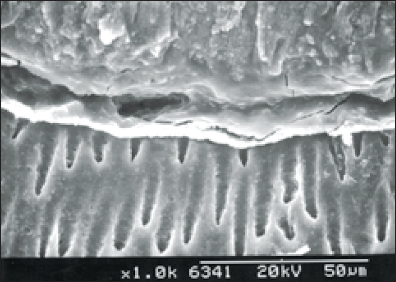
Figure 21. Photograph of the NaOCl-6 group Level 1 (× 1000); Scanning electron micrographs of tooth sections showing the gap-free region between gutta-percha and the surface of root canals. Surface generally covered with smear layer.
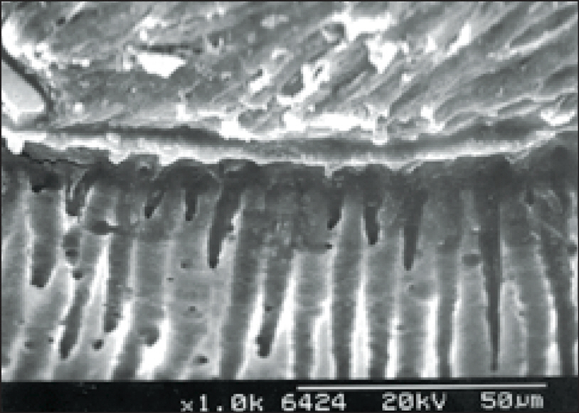
Figure 22. Photograph of the NaOCl-6 group Level 2 (× 1000); Scanning electron micrographs of tooth sections showing the gap-free region between gutta-percha and the surface of root canals. Surface generally covered with smear layer.
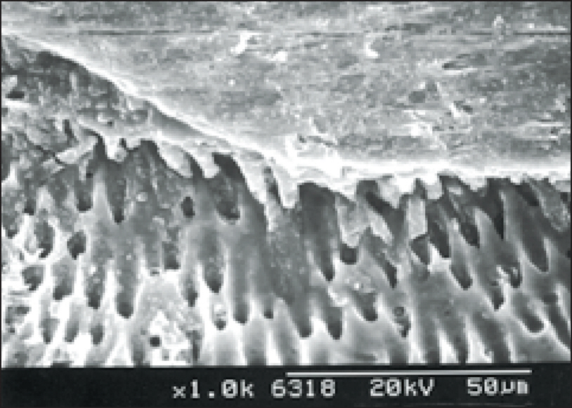
Figure 23. Photograph of the EDTA-6 group Level 1 (× 1000); Scanning electron micrographs of tooth sections showing the penetration of sealers to opened dentinal tubules. A few sealer tags could be seen within the intertubular dentin.
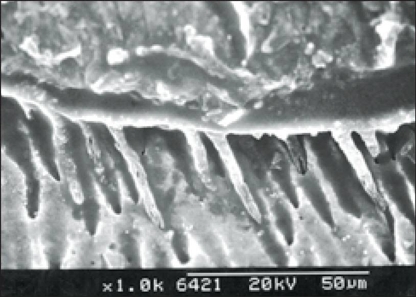
Figure 24. Photograph of the EDTA-6 group Level 2 (× 1000); Scanning electron micrographs of tooth sections showing the penetration of sealers to opened dentinal tubules. A few sealer tags could be seen within the intertubular dentin.
Cited by 2 articles
-
The effect of MTAD as a final root canal irrigants on the coronal bacterial leakage of obturated root canals
Tae Woo Kim, Seok Woo Chang, Dong Sung Park
J Korean Acad Conserv Dent. 2008;33(4):397-404. doi: 10.5395/JKACD.2008.33.4.397.Effect of soft chelating irrigation on the sealing ability of GP/AH Plus root fillings
Yi-Suk Yu, Tae-Gun Kim, Kwang-Won Lee, Mi-Kyung Yu
J Korean Acad Conserv Dent. 2009;34(6):484-490. doi: 10.5395/JKACD.2009.34.6.484.
Reference
-
참고문헌
1. Yamada RS, Armas A, Goldman M, Lin PS. A scanning electron microscopic comparison of a high volume flush with several irrigating solutions. Part 3. J Endod. 9(4):137–142. 1983.2. Gutmann JL, Witherspoon DE. Pathways of the pulp. 7th ed.Mosby Inc.;St. Louis, Mo: p. p272. 1998.3. Ingle JI, Backland LK. Endodontics. 5th ed. BC Decker Inc;Loma Linda, California: p. p503–505. 2002.4. Russell AD, Hugo WB, Ayliffe GAJ. Principles and Practice of Disinfection, Preservation, and Sterilization. 3rd ed. Blackwell Science;Malden, MA: p. p99–100. 1999.5. Calt S, Serper A. Time-dependent effects of EDTA on dentin structures. J Endod. 28(1):17–19. 2002.
Article6. Lee JK, Choi GW. Time-dependent effects of EDTA on removal of smear layer in the root canal system. J Kor Acad Cons Dent. 2006:(In Press).7. Cergneux M, Ciucchi B, Dietschi JM, Holz J. The influence of the smear layer on the sealing ability of canal obturatiion. Int Endod J. 20(5):228–232. 1987.8. White RR, Goldman M, Lin P. The influence of the smeared layer upon dentinal tubule penetration by plastic filling materials. J Endod. 10(12):558–562. 1984.
Article9. Drake D R, Wiemann A H, Rivera E M and, Walton R E. Bacterial retention in canal walls in vitro effect of smear layer. J Endod. 20(2):78–82. 1994.
Article10. Goldman LB, Goldman M, Kronman JH, Lin PS. The efficacy of several irrigating solutions for endodontics: A scanning electron microscopic study. Oral Surg Oral Med Oral Pathol. 52(2):197–204. 1981.
Article11. Walters MJ, Baumgartner JC, Marshall JG. Efficacy of Irrigation with Rotary Instrumentation. J Endod. 28(12):837–839. 2002.
Article12. Cunningham WT, Balekjian AY. Effect of temperature on collagen- dissolving ability of sodium hypochlorite endodontic irrigant. Oral surg Oral Med Oral Pathol. 49(2):175–177. 1980.13. Berutti E, Marini R. A Scanning Electron Microscopic Evaluation of the Debridement Capability of Sodium Hypochlorite at Different Temperatures. J Endod. 22(9):467–470. 1996.
Article14. Baumgartner JC, Mader CL. A scanning electron microscopic evaluation of four oot canal irrigation regimens. J Endod. 13(4):147–157. 1987.15. Czonstkowsky M, Michanowicz A, Vazquez JA. Evaluation of an injection of thermoplasticized low-temperature gutta-percha using radioactive isotopes. J Endod. 11(2):71–74. 1985.
Article16. Tucker JW, Mizrahi S, Seltzer S. Scanning electron microscopic study of the efficacy of various irrigating solutions: Urea, Tublicid Red, and Tubulicid Blue. J Endod. 2(3):71–78. 1976.17. Williams S, Goldman M. Penetrability of the smeared layer by strain of proteus vulgaris. J Endod. 11(9):385–388. 1985.18. Pashley DH. Smear layer: physiological considerations. Oper Dent Suppl. 3:13–29. 1984.19. Orstavik D, Haapasalo M. Disinfection by endodontic irrigants and dressings or experimentally infected dentinal tubules. Endod Dent Traumatol. 6(4):142–149. 1990.20. Love RM, Chandler NP, Jenkinson HF. Penetration of smeared or non smeared dentine by Streptococcus gor-donii. Int Endod J. 29(1):2–12. 1996.21. Ruddle CJ. Pathways of the pulp. 8th ed.Mosby Inc.;St. Louis, Mo: p. p258. 2002.22. Hand RE, Smith ML, Harrison JW. Analysis of the effect of dilution on the necrotic tissue dissolution property of sodium hypochlorite. J Endod. 4(2):60–64. 1978.
Article23. Patterson SS. In vivo and in vitro studies of the effect of the disodium salt of ethylenediamine tetra-acetate on human dentine and its endodontic implications. Oral surg Oral Med Oral Pathol. 16:83–103. 1963.
Article24. Goldman M, Goldman LB, Cavaleri R, Bogis J, Lin PS. The efficacy of several endodontic irrigating solutions: a scanning electron microscopic study: part 2. J Endod. 8(11):487–492. 1982.
Article25. Grawehr M, Sener B, Waltimo T, Zehnder M. Interactions of ethylenediamine tetraacetic acid with sodium hypochlorite in aqueous solutions. Int Endod J. 36(6):411–415. 2003.
Article26. Tamse A, Katz A and, Kablan F. Comparison of apical leakage shown by four different dyes with two evaluating methods. Int Endod J. 31(5):333–337. 1998.
Article27. Madison S, Krell KV. Comparison of EDTA and sodium hypochlirite on the apical seal of endodontically treated teeth. J Endod. 10(10):499–503. 1984.28. Saunders WP, Saunders EM. The effect of smear layer upon the coronal leakage of gutta-percha root fillings and a glass ionomer sealer. Int Endod J. 25(5):145–149. 1992.
Article29. Syngcuk Kim. Pathways of the pulp. 8th ed.Mosby Inc.;St. Louis, Mo: p. p707–708. 2002.
- Full Text Links
-
- Actions
-
Cited
- CITED
-
- Close
- Share
-
- Similar articles
-
- Effect of microleakage of a self-etching primer adhesive according to types of cutting instruments
- The comparison of microleakage on intracoronal restoration after non-vital bleaching
- Microleakage of composite resin restoration according to the number of thermocycling
- Time-dependent effects of EDTA application on removal of smear layer in the root canal system
- Microleakage and marginal hybrid layer of dentin adhesives
