J Korean Acad Conserv Dent.
2006 Mar;31(2):133-140. 10.5395/JKACD.2006.31.2.133.
Assessment of sterilization effect and the alteration of surface texture and physical properties of gutta-percha cone after short-term chemical disinfection
- Affiliations
-
- 1Department of Conservative Dentistry, Oral Science Research Center, Yonsei University, Korea.
- 2Department of Oral Biology, Oral Science Research Center, Yonsei University, Korea.
- 3Department of Conservative Dentistry, Dental Research Institute, Seoul National University, Korea. kum6139@snu.ac.kr
- KMID: 1986859
- DOI: http://doi.org/10.5395/JKACD.2006.31.2.133
Abstract
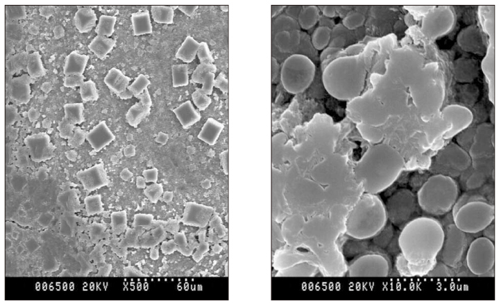
- The purposes of this study were firstly to identify the microbial species on gutta-percha (GP) cones exposed at clinics using polymerase chain reaction, and secondly to evaluate the short-term sterilization effect of three chemical disinfectants. It also evaluated the alteration of surface texture and physical properties of GP cones after 5-min soaking into three chemical disinfectants. 150 GP cones from two endodontic departments were randomly selected for microbial detection using PCR assay with universal primer. After inoculation on the sterilized GP cones with the same microorganism identified by PCR assay, they were soaked in three chemical disinfectants: 5% NaOCl, 2% Chlorhexidine, and ChloraPrep for 1, 5, 10, and 30 minutes. The sterilization effect was evaluated by turbidity and subculture. The change of surface textures using a scanning electron microscope and the tensile strength and elongation rate of the GP cones were measured using an Instron 5500 (Canton). Statistical analysis was performed. Four bacterial species were detected in 29 GP cones (19.4%), and all the species belonged to the genus Staphylococcus. All chemical disinfectants were effective in sterilization with just 1 minute soaking. On the SEM picture of NaOCl-soaked GP cone, a cluster of cuboidal crystals was seen on the cone surface. The tensile strength of NaOCl-soaked group was significantly higher than the other groups (p < 0.05). Also, all disinfectants significantly increased the elongation rate of GP cones compared to the fresh GP cone (p < 0.05). Present data demonstrate that three chemical disinfectants are useful for rapid sterilization of GP cone just before obturation.
MeSH Terms
Figure
Reference
-
1. Moorer WR, Genet JM. Evidence for antibacterial activity of endodontic gutta-percha cones. Oral Surg Oral Med Oral Pathol. 1982. 53:503–507.
Article2. Delivanis PD, Mattison GD, Mendel RW. The survivability of F43 strain of streptococcus sanguis in root filled with gutta-percha and Procosol cement. J Endod. 1983. 9:407–410.
Article3. Cardoso CL, Kotaka CR, Guilhermetti M, Hidalgo MM. Rapid sterilization of gutta-percha cones with glutaraldehyde. J Endod. 1998. 24:561–563.
Article4. Cardoso CL, Kotaka CR, Redmerski R, Guilhermetti M, Queiroz AF. Rapid decontamination of gutta-percha cones with sodium hypochlorite. J Endod. 1999. 25:498–501.
Article5. da Motta PG, de Figueiredo CB, Maltos SM, Nicoli JR, Ribeiro Sobrinho AP, Maltos KL, Carvalhais HP. Efficacy of chemical sterilization and storage conditions of gutta-percha cones. Int Endod J. 2001. 34:435–439.
Article6. de Souza RE, de Souza EA, Sousa-Neto MD, Pietro RC. In vitro evaluation of different chemical agents for the decontamination of gutta-percha cones. Pesqui Odontol Bras. 2003. 17:75–77.
Article7. Siqueira JF JR, da Siliva CH, Cerqueira M das D, Lopes HP, de Uzeda M. Effectiveness of four chemical solutions in eliminating Bacillus subtilius spores on gutta-percha cones. Endod Dent Traumatol. 1998. 14:124–126.
Article8. Möller B, Orstavik D. Influence of antiseptic storage solution on physical properties of endodontic gutta-percha points. Scand J Dent Res. 1985. 93:158–161.9. Lee MS. An experimental study of the effect of the various antiseptic storage solutions on physical properties of gutta-percha cone. 1989. Yonsei Dental College;MS Thesis.10. Siqueira JF JR, Rocas IN. PCR methodology as a valuable tool for identification of endodontic pathogens. J Dent. 2003. 31:333–339.
Article11. Gill SR, Fouts DE, Archer GL. Insights on evolution of virulence and resistance from the complete genome analysis of an early Mehicillin-Resistant Staphylococcus aureus strain and a biofilm-producing Methicillin-Resistant staphylococcus epidermidis strain. J Bacteriol. 2005. 187:2426–2438.
Article12. Noiri Y, Ehara A, Kawahara T, Takemura N, Ebisu S. Participation of bacterial biofilms in refractory and chronic periapical periodontitis. J Endod. 2002. 28:679–683.
Article13. Takemura N, Noiri Y, Ehara A, Kawahara T, Noguchi N, Ebuisu S. Single species biofilm-forming ability of root canal isolates on gutta-percha points. Eur J Oral Sci. 2004. 112:523–529.
Article14. Short RD, Dorn SO, Kuttler S. The crystallization of sodium hypochlorite on gutta-percha cones after the rapid-sterilization technique: an SEM study. J Endod. 2003. 29:670–673.
Article15. Valois CR, Silva LP, Azevedo RB. Effects of 2% chlorhexidine and 5.25% sodium hypochlorite on gutta-percha cones studied by atomic force microscopy. Int Endod J. 2005. 38:425–429.
Article
- Full Text Links
- Actions
-
Cited
- CITED
-
- Close
- Share
- Similar articles
-
- Obturation efficiency of non-standardized gutta-percha cone in curved root canals prepared with 0.06 taper nickel-titanium instruments
- A study of insertion depth of gutta percha cones after shaping by Ni-Ti rotary files in simulated canals
- A comparison of thermoplasticized injectable gutta-percha techniques in ribbon-shaped canals : adaptation to canal walls
- The effect of gutta-percha removal using nickel-titanium rotary instruments
- Assessment of vertical root fracture using cone-beam computed tomography


