J Clin Neurol.
2006 Dec;2(4):231-237. 10.3988/jcn.2006.2.4.231.
Preliminary Studies on the Clinical Features of Multiple Sclerosis in Korea
- Affiliations
-
- 1Department of Neurology, Chungbuk National University College of Medicine, Chungbuk, Korea. sslee@chungbuk.ac.kr
- 2Department of Neurology, Chungnam National University College of Medicine, Daejeon, Korea.
- 3Department of Neurology, Sun General Hospital, Daejeon, Korea.
- KMID: 1980462
- DOI: http://doi.org/10.3988/jcn.2006.2.4.231
Abstract
- BACKGROUND AND PURPOSE
Multiple sclerosis (MS) in Asians is characterized by frequent involvement of the spinal cord and optic nerve and low prevalence rates, but even the most fundamental epidemiologic findings and unique clinical features of MS patients in Korea have not been studied extensively. We performed this study to establish the clinical spectrum of MS patients in Korea.
METHODS
Sixty-two MS patients (25 men and 37 women) who satisfied the diagnostic criteria for definite MS were reviewed retrospectively using medical records from two university hospitals and one general hospital. The MS patients were classified into the three clinical subtypes according to the involved site (opticospinal, spinal, and conventional MS).
RESULTS
The age at MS onset was 35.2+/-13.3 (mean+/-SD) years, and the predominant initial clinical manifestations were myelopathy (54.8%) and optic neuropathy (33.9%). The single most common involved lesion site was the spinal cord (35.5%). Spinal (35.5%) and opticospinal (25.8%) MS were the most common type, and they had a frequent relapsing-remitting course and long lesions extending over two vertebral segments (as assessed using spinal cord MRI). The interval between the first symptom and relapse was 35.6+/-71.1 months, and the number of relapses was 3.8+/-2.6. The spinal form of MS was associated with a higher age at onset and a higher male-to-female ratio than the other types. Positive rates of CSF oligoclonal bands and IgG index and the number of patients with characteristic brain MRI lesions were low. However, the abnormal rate of visual evoked potentials was relatively high (64.4%).
CONCLUSION
The clinical features of MS patients in Korea are different from those in Western patients, but similar to those in Far East Asian patients. The value of the various diagnostic tools used for MS should therefore be reevaluated, at least for Korean patients.
MeSH Terms
-
Asian Continental Ancestry Group
Brain
Evoked Potentials, Visual
Far East
Hospitals, General
Hospitals, University
Humans
Immunoglobulin G
Korea*
Magnetic Resonance Imaging
Male
Medical Records
Multiple Sclerosis*
Oligoclonal Bands
Optic Nerve
Optic Nerve Diseases
Optic Neuritis
Prevalence
Recurrence
Retrospective Studies
Spinal Cord
Spinal Cord Diseases
Immunoglobulin G
Oligoclonal Bands
Figure
-
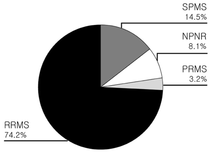
Figure 1 Clinical courses of MS: RRMS; relapsing-remitting MS, SPMS; secondary progressive MS, PRMS; progressive relapsing MS, NPNR; nonprogressing and nonrelapsing MS
Reference
-
1. Mayr WT, Pittock SJ, McClelland RL, Jorgensen NW, Noseworthy JH, Rodriguez M. Incidence and prevalence of multiple sclerosis in Olmsted County, Minnesota, 1985-2000. Neurology. 2003. 61:1373–1377.
Article2. Ropper AH, Brown RH. Adams and Victor's principles of neurology. 2005. 8th ed. New York: McGraw-Hill;771–796.3. Kuroiwa Y, Igata A, Itahara K, Koshijima S, Tsubaki T. Nationwide survey of multiple sclerosis in Japan. Clinical analysis of 1,084 cases. Neurology. 1975. 25:845–851.
Article4. Yamasaki K, Horiuchi I, Minohara M, Kawano Y, Ohyagi Y, Yamada T, et al. HLA-DPB10531-associated opticospinal multiple sclerosis. Clinical, neuroimaging and immunogenetic studies. Brain. 1999. 122:1689–1696.
Article5. Kira J. Multiple sclerosis in the Japanese population. Lancet Neurol. 2003. 2:117–127.
Article6. Cho YJ, Jeon BS, Kim YH, Chang KH. Clinical features and outcomes from diagnostic work-up in definite multiple sclerosis. J Korean Neurol Assoc. 1999. 17:823–828.7. Poser CM, Paty DW, Scheinberg L, McDonald WI, Davis FA, Ebers GC, et al. New diagnostic criteria for multiple sclerosis: guidelines for research protocols. Ann Neurol. 1983. 13:227–231.
Article8. McDonald WI, Compston A, Edan G, Goodkin D, Hartnung H, Lublin FD, et al. Recommended diagnostic criteria for multiple sclerosis: guidelines from the international panel on the diagnosis of multiple sclerosis. Ann Neurol. 2001. 50:121–127.
Article9. Houzen H, Niino M, Kikuchi S, Fukazawa T, Nogoshi S, Matsumoto H, et al. The prevalence and clinical characteristics of MS in northern Japan. J Neurol Sci. 2003. 211:49–53.
Article10. Jacobs LD, Wende KE, Brownscheidle CM, Apatoff B, Coyle PK, Goodman A, et al. A profile of multiple sclerosis: the New York State Multiple Sclerosis Consortium. Mult Scler. 1999. 5:369–376.
Article11. Kim YH, Chang KH, Kim SS, Park BK, Seong CK, Han MH, et al. Multiple sclerosis of the spinal cord: MR imaging findings. J Korean Radiol Soc. 1988. 39:427–433.
Article12. Thielen KR, Miller GM. Multiple sclerosis of the spinal cord: Magnetic resonance appearance. J Comput Assist Tomogr. 1996. 20:434–438.
Article13. Nakashima I, Fujihara K, Miyazawa H, Misu T, Fujimori J, Sato S, et al. Relevance of callosal and periventricular MRI lesions to oligoclonal bands in multiple sclerosis. Acta Neurol Scand. 2006. 113:125–131.
Article14. Lennon VA, Wingerchuk DM, Kryzer TJ, Pittock SJ, Lucchinetti CF, Fujihara K, et al. A serum autoantibody marker of neuromyelitis optica: distinction from multiple sclerosis. Lancet. 2004. 364:2106–2112.
Article15. Kikuchi S, Fukazawa T. "OSMS is NMO, but not MS": confirmed by NMO-IgG? Lancet Neurol. 2005. 4:594–595.
Article
- Full Text Links
-
- Actions
-
Cited
- CITED
-
- Close
- Share
-
- Similar articles
-
- Understanding MRI Features of Multiple Sclerosis: Based on the 2017 McDonald Criteria
- The Effect of High-dose Intravenous Steroid("pulse") Therapy in Neurologic Disease-Preliminary Report
- Multiple Sclerosis in Korea: Its Clinical Features and Estimated Prevalence Rate
- Clinical Features and Prognosis of Optic Neuritis: A Preliminary Report
- Manifestations Like Multiple Sclerosis in a Paitent with Rheumatoid Arthritis and Sjogren's Syndrome
