J Cardiovasc Ultrasound.
2014 Dec;22(4):230-231. 10.4250/jcu.2014.22.4.230.
Valve in Valve: Three-Dimensional Transoesophageal Echocardiogram and Multi-Slice CT Images of Bio-Prosthetic Aortic Valve Replaced by Medtronic CoreValve
- Affiliations
-
- 1Department of Cardiology and Cardiothoracic Surgery, Chesterman Wing, Northern General Hospital, Sheffield Teaching Hospitals NHS Foundation Trust, Sheffield, UK. pankajvic@gmail.com
- KMID: 1980430
- DOI: http://doi.org/10.4250/jcu.2014.22.4.230
Abstract
- No abstract available.
Keyword
MeSH Terms
Figure
-

Fig. 1 Three-dimensional transoesophageal view of Medtronic CoreValve (blue arrows) embedded in sinus of Valsalva, where bio-prosthetic aortic valve was previously implanted. In this systolic view the CoreValve cusps are open (red arrows). LA: left atrium, LV: left ventricle, LVOT: left ventricular outflow tract.
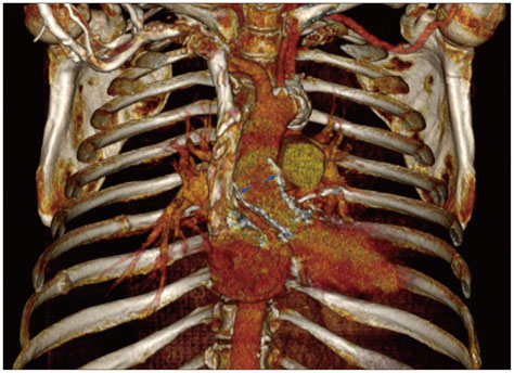
Fig. 2 Computed tomography three-dimensional multi-slice reconstruction showing well-seated CoreValve strut (blue arrows) in the left ventricular out-flow tract to aortic root.
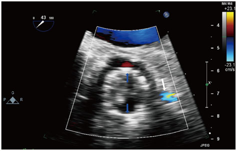
Fig. 3 Colour Doppler view in two-dimensional short-axis transoesophageal aortic valve plane showing the strut of Medtronic CoreValve (blue arrows) and demonstrating clear flow in the left main stem (white arrow).
Reference
-
1. Eggebrecht H, Schäfer U, Treede H, Boekstegers P, Babin-Ebell J, Ferrari M, Möllmann H, Baumgartner H, Carrel T, Kahlert P, Lange P, Walther T, Erbel R, Mehta RH, Thielmann M. Valve-in-valve transcatheter aortic valve implantation for degenerated bioprosthetic heart valves. JACC Cardiovasc Interv. 2011; 4:1218–1227.
Article
- Full Text Links
-
- Actions
-
Cited
- CITED
-
- Close
- Share
-
- Similar articles
-
- Echocardiographic Preoperative Prediction of Prosthetic Aortic Valve Size in Patient with Aortic Valve Replacment
- A Case of Double Orifice Mitral Valve in a Patient with Bicuspid Aortic Valve: Coincidental or a Missed Finding?
- Surgical Management of Aortic Valve Injury after Nonpenetrating Trauma
- Quadricuspid Aortic Valve : Report of Three Cases and Review of the Literature
- A Case of Prosthetic Valve Dysfunction in the Aortic Position: Caused by Pannus Formation
