Spontaneously Healed Membranous Type Ventricular Septal Defect with Malaligned Interventricular Septal Wall and Double-Chambered Right Ventricle in a 56-Year-Old Patient
- Affiliations
-
- 1Department of Internal Medicine, School of Medicine, The Catholic University of Korea, Seoul, Korea. younhj@catholic.ac.kr
- KMID: 1980370
- DOI: http://doi.org/10.4250/jcu.2011.19.3.148
Abstract
- A 56-year-old male presented with resting dyspnea and chest discomfort for several years. During transthoracic and transesophageal echocardiography, a spontaneously healed membranous type ventricular septal defect (VSD) with malaligned interventricular septal wall, aneurysmal changes, a subaortic ridge and a double-chambered right ventricle (DCRV) was observed. When combined with DCRV, VSD with malalignment between the outlet and trabecular septa was associated with tetralogy of Fallot. The subaortic ridge was due to turbulent flow caused by the malalignment-type VSD. The VSD with malaligned interventricular septal wall can be developed after aneurismal changes of a perimembranous VSD. We report here in the unusual case of a 56-year-old patient who had a pathology complex comprising DCRV, subaortic ridge, spontaneously healed membranous type VSD with malaligned interventricular septal wall, and survived with surgical treatment.
Keyword
MeSH Terms
Figure
-
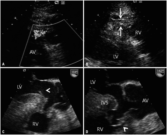
Fig. 1 Preoperative transthoracic and transesophageal echocardiography. (A) Transthoracic echocardiography reveals a high-velocity systolic jet arising in the mid right ventricular outflow tract. (B) Right ventricular hypertrophy is seen on the parasternal long axis view. Transesophageal echocardiography reveals a subaortic ridge (arrowhead) (C) and aneurysmal changes (arrowhead) of the ventricular septal defect with anterior deviated interventricular septum (D). AV: aortic valve, RV: right ventricle, LV: left ventricle, IVS: interventricular septum.
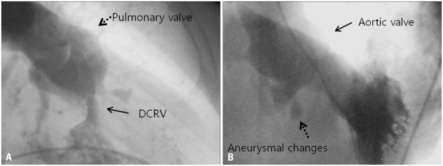
Fig. 2 Preoperative right and left ventriculography. Right ventriculography at end-systole reveals an anomalous muscle bundle dividing the cavity into 2 chambers (A), and left ventriculography shows spontaneous closed malalignmented ventricular septal defect with aneurysmal changes (B). DCRV: double-chambered right ventricle.
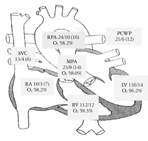
Fig. 3 Preoperative cardiac catheterization demonstrates a right ventricle-to-pulmonary artery gradient of 89 mmHg. There is no significant step-up of O2 saturation in the right ventricular sample that is higher than the highest right atrial sample because of spontaneously closed malalignmented ventricular septal defect. Systolic pressure/diastolic pressure is expressed as a mean pressure and O2 saturation as a percentage. SVC: superior vena cava, RA: right atrium, RV: right ventricle, MPA: main pulmonary artery, RPA: right pulmonary artery, PCWP: pulmonary capillary wedge pressure, LV: left ventricle.
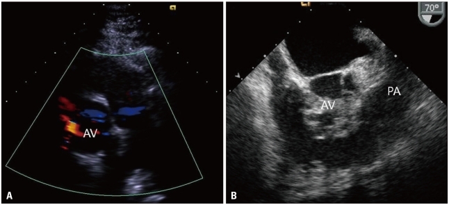
Fig. 4 Postoperative transthoracic and transesophageal echocardiography. A: Transthoracic echocardiography reveals that a high-velocity systolic jet arising in the mid right ventricular outflow tract disappeared after surgery. B: Transesophageal echocardiography reveals much improved right ventricular outflow tract diameter after surgery. AV: aortic valve, PA: pulmonary artery.
Reference
-
1. Maron BJ, Ferrans VJ, White RI Jr. Unusual evolution of acquired infundibular stenosis in patients with ventricular septal defect. Clinical and morphologic observations. Circulation. 1973; 48:1092–1103.2. Choi YJ, Park SW. Characteristics of double-chambered right ventricle in adult patients. Korean J Intern Med. 2010; 25:147–153. PMID: 20526387.3. McElhinney DB, Chatterjee KM, Reddy VM. Double-chambered right ventricle presenting in adulthood. Ann Thorac Surg. 2000; 70:124–127. PMID: 10921695.4. Wu MH, Wu JM, Chang CI, Wang JK, Wu YN, Chien SC, Lue HC. Implication of aneurysmal transformation in isolated perimembranous ventricular septal defect. Am J Cardiol. 1993; 72:596–601. PMID: 8362777.5. Matina D, van Doesburg NH, Fouron JC, Guérin R, Davignon A. Subxiphoid two-dimensional echocardiographic diagnosis of double-chambered right ventricle. Circulation. 1983; 67:885–888. PMID: 6825244.6. Wang JK, Wu MH, Chang CI, Chiu IS, Chu SH, Hung CR, Lue HC. Malalignment-type ventricular septal defect in double-chambered right ventricle. Am J Cardiol. 1996; 77:839–842. PMID: 8623736.7. Zielinsky P, Rossi M, Haertel JC, Vitola D, Lucchese FA, Rodrigues R. Subaortic fibrous ridge and ventricular septal defect: role of septal malalignment. Circulation. 1987; 75:1124–1129. PMID: 3568324.8. Wu MH, Wang JK, Chang CI, Chiu IS, Lue HC. Implication of anterior septal malalignment in isolated ventricular septal defect. Br Heart J. 1995; 74:180–185. PMID: 7546999.9. Oppenheimer-Dekker A, Gittenberger-de Groot AC, Bartelings MM, Wenink AC, Moene RJ, van der Harten JJ. Abnormal architecture of the ventricles in hearts with an overriding aortic valve and a perimembranous ventricular septal defect ("Eisenmenger VSD"). Int J Cardiol. 1985; 9:341–355. PMID: 4055151.10. Vogel M, Smallhorn JF, Freedom RM, Coles J, Williams WG, Trusler GA. An echocardiographic study of the association of ventricular septal defect and right ventricular muscle bundles with a fixed subaortic abnormality. Am J Cardiol. 1988; 61:857–860. PMID: 3354451.11. Kim CJ, Chai IH, Koh KK, Sohn DW, Lee MM, Park YB, Choi YS, Seo JD, Lee YW. Double chambered right ventricle (DCRV) in adult and adolescence. Korean Circ J. 1990; 20:248–255.
- Full Text Links
-
- Actions
-
Cited
- CITED
-
- Close
- Share
-
- Similar articles
-
- A Case of Double Chambered Right Ventricle with Congenital Right Ventricular True Diverticulum
- Two Cases of Double-Chambered Right Ventricle without Other Congenital Cardiac Anomalies
- Two-chambered right ventricle resulting from aberrant muscle bundles a case report
- Double Chambered Right Ventricle with Ventricular Septal Defect in Adults: Case Series and Review of the Literature
- Noonan Syndrome with Double-Chambered Right Ventricle and Atrial Septal Defect: 1 Case Report
