Carcinoid Heart Disease: A Rare Cause of Right Ventricular Dysfunction Evaluation by Transthoracic 2D, Doppler and 3-D Echocardiography
- Affiliations
-
- 1Department of Internal Medicine, Faculty of Medicine, Geriatry-Second University of Naples (S.U.N.), Naples, Italy. federico.cacciapuoti@unina2.it
- 2Montevergine Cardiology Clinic (Avellino), Mercogliano, Italy.
- KMID: 1980360
- DOI: http://doi.org/10.4250/jcu.2011.19.2.99
Abstract
- Carcinoid heart disease is a rare cause of heart failure with or without right valvular heart impairments. In this study, we showed a case of carcinoid tumour with hepatic metastases inducing carcinoid heart disease. Neuroendocrine heart involvement happens for severe tricuspid valve insufficiency and plaques on right ventricular (RV) walls produced by a release of serotonin (5-HT). A patient affected by primitive ileal tumour with 5-HT-secernent hepatic metastases inducing tricuspid insufficiency is showed. Transthoracic 2-D echocardiography showed tricuspid valve regurgitation and both right atrium, RV-walls plaques and RV dilation. Continue-wave Doppler showed a characteristic "dagger shaped" spectrum of tricuspid systolic flow. RV function was evaluated with 3-D transthoracic echocardiography. In particular, RV volumes, RV ejection fraction and stroke volume were defined by this technique. 2, 3-D echocardiography and Doppler method are useful techniques to show heart valves' derangements and RV function to non-invasively detect RV impairments in carcinoid heart disease.
Keyword
MeSH Terms
Figure
-
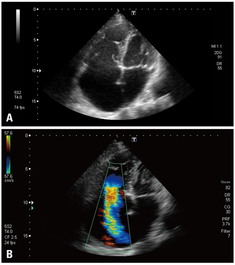
Fig. 1 A: Two-dimensional echocardiography recorded in the patient with carcinoid heart disease. Dilatation of right cavities and thickened, fixed and retracted tricuspid leaflets. Endocardial plaques on right ventricular walls are also evident. B: Severe tricuspid regurgitation seen in the same patient at color flow imaging.
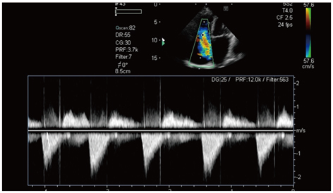
Fig. 2 "Dagger shaped" of systolic tricuspid flow, characterized by an early peak pressure and its rapid decline.
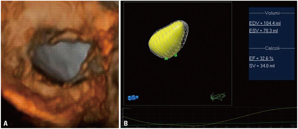
Fig. 3 A: Three-dimensional transthoracic echocardiography showing the adhesion of tricuspid leaflets to right ventricular walls, that prevents the valve closure during systole. B: Three-dimensional shape of diastolic and systolic volumes (EDV and ESV), ejection fraction% (EF%) and stroke volume (SV) of right ventricle.
Reference
-
1. Kulke MH, Mayer RJ. Carcinoid tumors. N Engl J Med. 1999. 340:858–868.
Article2. Ross EM, Roberts WC. The carcinoid syndrome: comparison of 21 necropsy subjects with carcinoid heart disease to 15 necropsy subjects without carcinoid heart disease. Am J Med. 1985. 79:339–354.
Article3. Bhattacharyya S, Davar J, Dreyfus G, Caplin ME. Carcinoid heart disease. Circulation. 2007. 116:2860–2865.
Article4. Waltenberger J, Lundin L, Oberg K, Wilander E, Miyazono K, Heldin CH, Funa K. Involvement of transforming growth factor-beta in the formation of fibrotic lesions in carcinoid heart disease. Am J Pathol. 1993. 142:71–78.5. Jian B, Xu J, Connolly J, Savani RC, Narula N, Liang B, Levy RJ. Serotonin mechanisms in heart valve disease I: serotonin-induced up-regulation of transforming growth factor-beta1 via G-protein signal transduction in aortic valve interstitial cells. Am J Pathol. 2002. 161:2111–2121.6. Soga J, Yakuwa Y, Osaka M. Carcinoid syndrome: a statistical evaluation of 748 reported cases. J Exp Clin Cancer Res. 1999. 18:133–141.7. Pellikka PA, Tajik AJ, Khandheria BK, Seward JB, Callahan JA, Pitot HC, Kvols LK. Carcinoid heart disease. Clinical and echocardiographic spectrum in 74 patients. Circulation. 1993. 87:1188–1196.
Article8. Tamborini G, Marsan NA, Gripari P, Maffessanti F, Brusoni D, Muratori M, Caiani EG, Fiorentini C, Pepi M. Reference values for right ventricular volumes and ejection fraction with real-time three-dimensional echocardiography: evaluation in a large series of normal subjects. J Am Soc Echocardiogr. 2010. 23:109–115.
Article9. Kjaergaard J, Petersen CL, Kjaer A, Schaadt BK, Oh JK, Hassager C. Evaluation of right ventricular volume and function by 2D and 3D echocardiography compared to MRI. Eur J Echocardiogr. 2006. 7:430–438.
Article10. De Simone R, Wolf I, Mottl-Link S, Böttiger BW, Rauch H, Meinzer HP, Hagl S. Intraoperative assessment of right ventricular volume and function. Eur J Cardiothorac Surg. 2005. 27:988–993.
Article11. Bhattacharyya S, Toumpanakis C, Burke M, Taylor AM, Caplin ME, Davar J. Features of carcinoid heart disease identified by 2- and 3-dimensional echocardiography and cardiac MRI. Circ Cardiovasc Imaging. 2010. 3:103–111.
Article
- Full Text Links
-
- Actions
-
Cited
- CITED
-
- Close
- Share
-
- Similar articles
-
- Echocardiographic Findings of Heart Disease in Children
- A Case of Coronary Artery-Left Ventricular Microfistulae Demonstrated by Transthoracic Doppler Echocardiography
- A Measurement of Pulmonary Flow, Systemic Flow and the Ratio of Pulmonary Flow and Systemic Flow by 2D-Doppler Echocardiography in Ventricular Septal Defect: A Comparison Study with the Fick's Method by Cardiac Catheterization
- The Usefulness of Doppler Tissue Image in Evaluation of Left Ventricular Systolic and Diastolic Dysfunction
- Functional Assessment for Congenital Heart Disease
