Severe bidentoalveolar protrusion treated with lingual Biocreative therapy using palatal miniplate
- Affiliations
-
- 1President of the Korean Society of Speedy Orthodontics, Korea.
- 2Division of Periodontology, Department of Dentistry, National Medical Center of Korea, Korea.
- 3Department of Orthodontics, Newyork University, NY, USA.
- 4Department of Orthodontics, College of Dentistry, Kyung Hee University, Korea. bravortho@hanmail.net
- 5Division of Orthodontics, Department of Orofacial Science, University of California San Francisco, CA, USA.
- KMID: 1975608
- DOI: http://doi.org/10.4041/kjod.2010.40.4.276
Abstract
- This case report describes the treatment of a 23-year, 8-month-old female patient with a Class II malocclusion who showed severe bidentoalveolar protrusion and anterior crowding. The treatment plan consisted of extracting all the first premolars, decrowding and en masse retraction of the upper six anterior teeth and lower anteriors. The upper C-plate placed in the midpalatal area combined with lingual sheath fixtures were used as substitutes for posterior anchorage teeth during upper anterior retraction. Preadjusted brackets (0.022-inch) were used for upper anterior decrowding. A 0.9 mm diameter stainless steel lever-arm soldered to the main arch wire facilitated controlled retraction of upper anteriors. The upper and lower dentition was detailed using a tooth positioner during the finishing stage. Correct overbite and overjet were obtained by decrowding and retraction of the upper six anterior teeth into their proper positions. Use of the C-plate and lingual appliances provided ideal anchorage to enhance the improvement in facial balance. The active treatment period was 19 months. The treatment result was stable 13 months after debonding.
Keyword
MeSH Terms
Figure
-
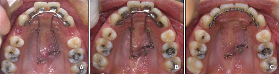
Fig. 1 Treatment progress showing occlusal photos of C-lingual retractor and C-plate combined retraction. A, One week after force application; B, 5 months after force application; C, decrowding and finishing using 2-D lingual brackets.
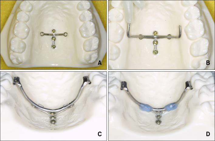
Fig. 2 Clinical procedure of C plate combined lingual tube appliance. A, C-plate inserted in the midpalatal suture area; B, application of 0.9 mm diameter stainless steel archwire for extension arm; C, 0.9 mm diameter lingual tubes are soldered to the extension arm and adapted to the C plate arm; D, resin application to the extension arm and C plate arm.
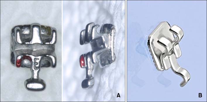
Fig. 3 Bracket systems for C plate combined retraction mechanics. A, 0.022-in slot Delta force low friction system (left, frontal view; right, lateral view); B, 2-D lingual bracket system.
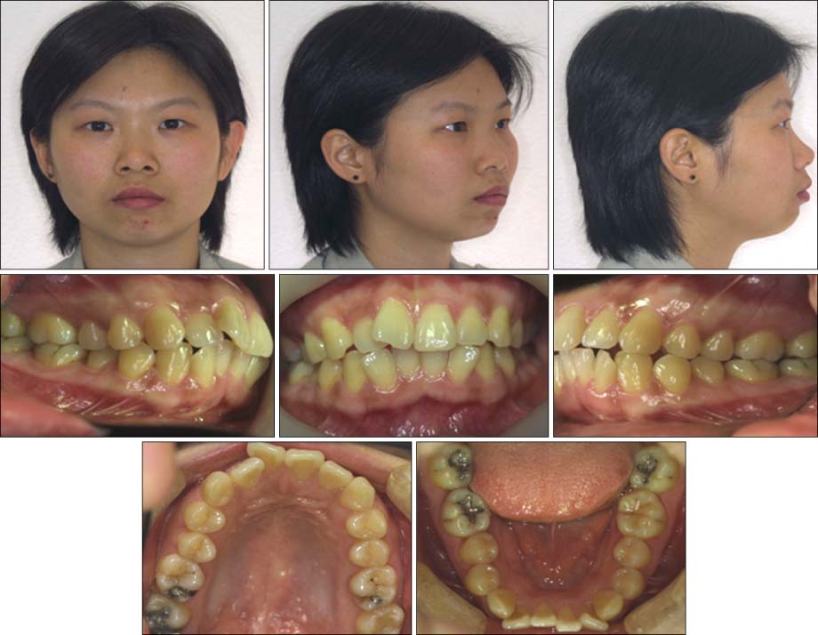
Fig. 4 Pretreatment extraoral and intraoral photographs.
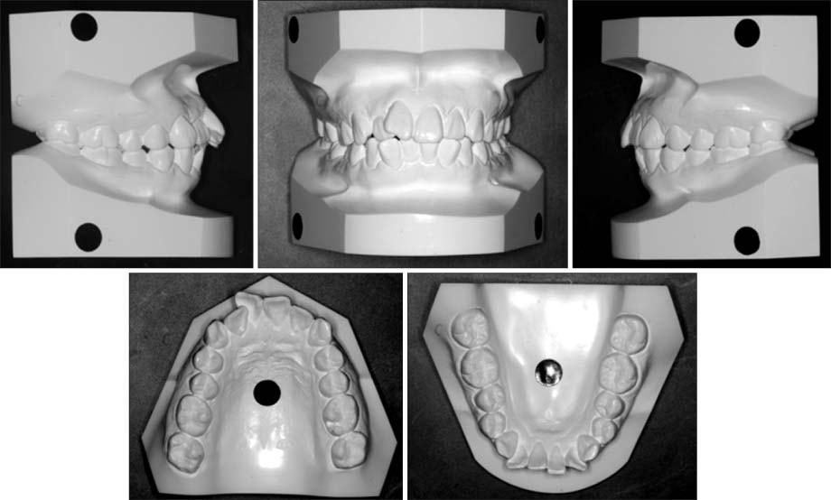
Fig. 5 Pretreatment study models.
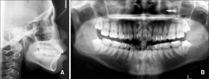
Fig. 6 Pretreatment lateral cephalometric radiograph (A) and panoramic radiograph (B).

Fig. 7 Progress intraoral photographs (A to E) and lateral cephalometric radiograph (F). En masse retraction of 6 anterior teeth with 0.016 × 0.022-inch stainless steel mushroom arch wire inserted in the C-plate combined lingual tube appliance hole, with power chains placed between the extended wire of the hole and cuspid bracket.
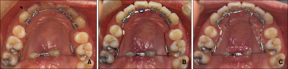
Fig. 8 Occlusal changes during the C-plate combined appliance dependent en masse retraction period. A, 4 months; B, 7 months; C, 12 months after force application.
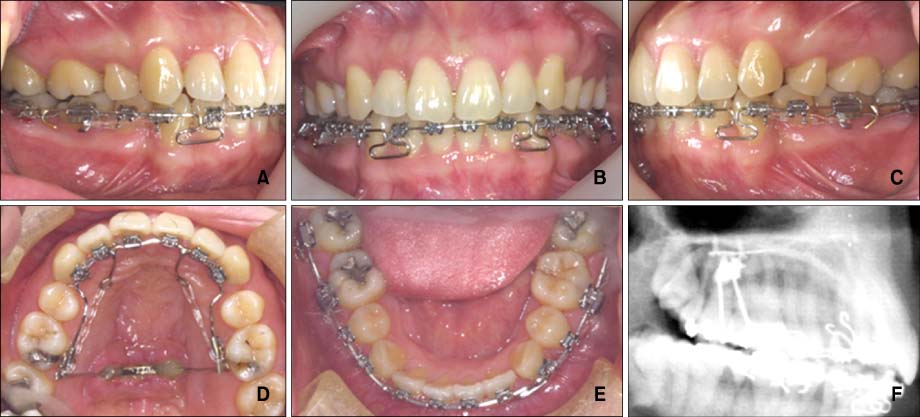
Fig. 9 Intraoral photographs (A to E) and lateral cephalometric radiograph (F) after 13 months of active treatment.
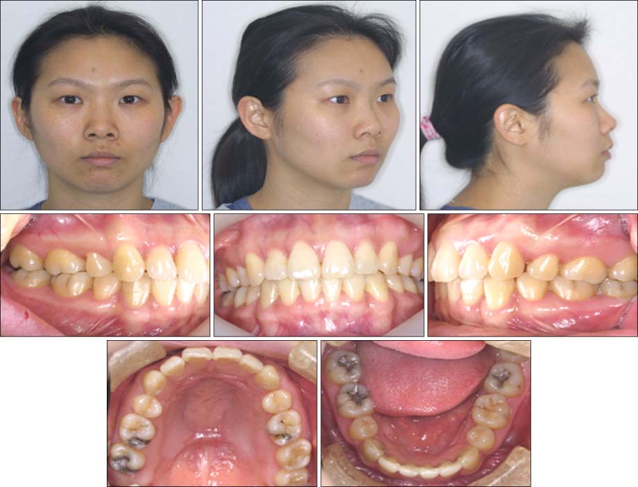
Fig. 10 Posttreatment extraoral and intraoral photographs.

Fig. 11 Posttreatment lateral cephalometric radiograph (A) and panoramic radiograph (B).
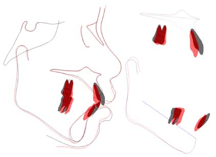
Fig. 12 Superimpositions of lateral cephalometric radiographs: pretreatment (black line) to posttreatment (red line).
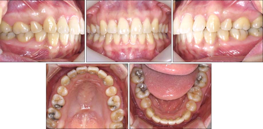
Fig. 13 13-month postretention intraoral photographs.
Cited by 2 articles
-
Torque control during lingual anterior retraction without posterior appliances
Sung-Seo Mo, Seong-Hun Kim, Sang-Jin Sung, Kyu-Rhim Chung, Yun-Sic Chun, Yoon-Ah Kook, Gerald Nelson
Korean J Orthod. 2013;43(1):3-14. doi: 10.4041/kjod.2013.43.1.3.Type of tooth movement during en masse retraction of the maxillary anterior teeth using labial versus lingual biocreative therapy in adults: A randomized clinical trial
Mais M. Sadek, Noha E. Sabet, Islam T. Hassan
Korean J Orthod. 2019;49(6):381-392. doi: 10.4041/kjod.2019.49.6.381.
Reference
-
1. Fulmer DT, Kuftinec MM. Cephalometric appraisal of patients treated with fixed lingual orthodontic appliances: historic review and analysis of cases. Am J Orthod Dentofacial Orthop. 1989. 95:514–520.
Article2. Miyawaki S, Yasuhara M, Koh Y. Discomfort caused by bonded lingual orthodontic appliances in adult patients as examined by retrospective questionnaire. Am J Orthod Dentofacial Orthop. 1999. 115:83–88.
Article3. Kurz C. The use of lingual appliances for correction of bimaxillary protrusion (four premolars extraction). Am J Orthod Dentofacial Orthop. 1997. 112:357–363.
Article4. Caniklioglu C, Oztürk Y. Patient discomfort: a comparison between lingual and labial fixed appliances. Angle Orthod. 2005. 75:86–91.5. Kim S, Park Y, Chung K. Severe anterior open bite malocclusion with multiple odontoma treated by C-lingual retractor and horseshoe mechanics. Angle Orthod. 2003. 73:206–212.6. Chung KR, Kook YA, Kim SH, Mo SS, Jung JA. Class II malocclusion treated by combining a lingual retractor and a palatal plate. Am J Orthod Dentofacial Orthop. 2008. 133:112–123.
Article7. Kim SH, Lee KB, Chung KR, Nelson G, Kim TW. Severe bimaxillary protrusion with adult periodontitis treated by corticotomy and compression osteogenesis. Korean J Orthod. 2009. 39:54–65.
Article8. Kim SH, Hwang YS, Ferreira A, Chung KR. Analysis of temporary skeletal anchorage devices used for en masse retraction: a preliminary study. Am J Orthod Dentofacial Orthop. 2009. 136:268–276.
Article9. Lim SM, Hong RK. Distal movement of maxillary molars using a lever-arm and mini-implant system. Angle Orthod. 2008. 78:167–175.
Article10. Umemori M, Sugawara J, Mitani H, Nagasaka H, Kawamura H. Skeletal anchorage system for open-bite correction. Am J Orthod Dentofacial Orthop. 1999. 115:166–174.
Article11. Kyung SH, Choi JH, Park YC. Miniscrew anchorage used to protract lower second molars into first molar extraction sites. J Clin Orthod. 2003. 37:575–579.12. Moon CH, Lee JS, Lee HS, Choi JH. Non-surgical treatment and retention of open bite in adult patients with orthodontic mini-implants. Korean J Orthod. 2009. 39:402–419.
Article13. Papadopoulos MA. Orthodontic treatment of Class II malocclusion with miniscrew implants. Am J Orthod Dentofacial Orthop. 2008. 134:604.e1. 604.e16.
Article14. Deguchi T, Murakami T, Kuroda S, Yabuuchi T, Kamioka H, Takano-Yamamoto T. Comparison of the intrusion effects on the maxillary incisors between implant anchorage and J-hook headgear. Am J Orthod Dentofacial Orthop. 2008. 133:654–660.
Article15. Lai EH, Yao CC, Chang JZ, Chen I, Chen YJ. Three-dimensional dental model analysis of treatment outcomes for protrusive maxillary dentition: comparison of headgear, miniscrew, and miniplate skeletal anchorage. Am J Orthod Dentofacial Orthop. 2008. 134:636–645.
Article16. Chaushu S, Becker A, Chaushu G. Lingual orthodontic treatment and absolute anchorage to correct an impacted maxillary canine in an adult. Am J Orthod Dentofacial Orthop. 2008. 134:811–819.
Article17. Park YC, Choi YJ, Choi NC, Lee JS. Esthetic segmental retraction of maxillary anterior teeth with a palatal appliance and orthodontic mini-implants. Am J Orthod Dentofacial Orthop. 2007. 131:537–544.
Article18. Chen YJ, Chang HH, Huang CY, Hung HC, Lai EH, Yao CC. A retrospective analysis of the failure rate of three different orthodontic skeletal anchorage systems. Clin Oral Implants Res. 2007. 18:768–775. Epub 2007 Sep 14.
Article19. Kang S, Lee SJ, Ahn SJ, Heo MS, Kim TW. Bone thickness of the palate for orthodontic mini-implant anchorage in adults. Am J Orthod Dentofacial Orthop. 2007. 131:4 Suppl. S74–S81.
Article20. Kravitz ND, Kusnoto B. Risks and complications of orthodontic miniscrews. Am J Orthod Dentofacial Orthop. 2007. 131:4 Suppl. S43–S51.
Article21. Miyawaki S, Koyana I, Inoue M, Mishima K, Sugahara T, Takano-Yamamoto T. Factors associated with the stability of titanium screws placed in the posterior region for orthodontic anchorage. Am J Orthod Dentofacial Orthop. 2003. 124:373–378.
Article
- Full Text Links
-
- Actions
-
Cited
- CITED
-
- Close
- Share
-
- Similar articles
-
- Type of tooth movement during en masse retraction of the maxillary anterior teeth using labial versus lingual biocreative therapy in adults: A randomized clinical trial
- Torque control during lingual anterior retraction without posterior appliances
- A Case of Lingual Myoclonus
- A cone-beam computed tomography study on strategic uprighting of mandibular molars using a biocreative reverse curve system
- Clinical application of maxillary tissue bone-borne expander and biocreative reverse curve system in the orthodontic retreatment of severe anterior open bite with transverse discrepancy: A case report
