Korean J Orthod.
2011 Jun;41(3):174-183. 10.4041/kjod.2011.41.3.174.
Factors affecting orthodontically induced root resorption of maxillary central incisors in the Korean population
- Affiliations
-
- 1Department of Orthodontics, School of Dentistry, Dankook University, Korea. abeh@dankook.ac.kr
- 2Department of Orthodontics, School of Dentistry, Kyunghee University, Korea.
- 3Okims Dental Office, Korea.
- KMID: 1975429
- DOI: http://doi.org/10.4041/kjod.2011.41.3.174
Abstract
OBJECTIVE
Orthodontically induced root resorption (OIRR) involves partial loss of cementum and dentin of teeth caused by routine orthodontic treatment. It decreases root length and influences the function of affected teeth. In this study, the treatment and patient factors causing apical root resorption in Koreans were determined. The observed factors were extraction, gender, age, displacement of root apex, total treatment period, total teeth length, and shape of the root.
METHODS
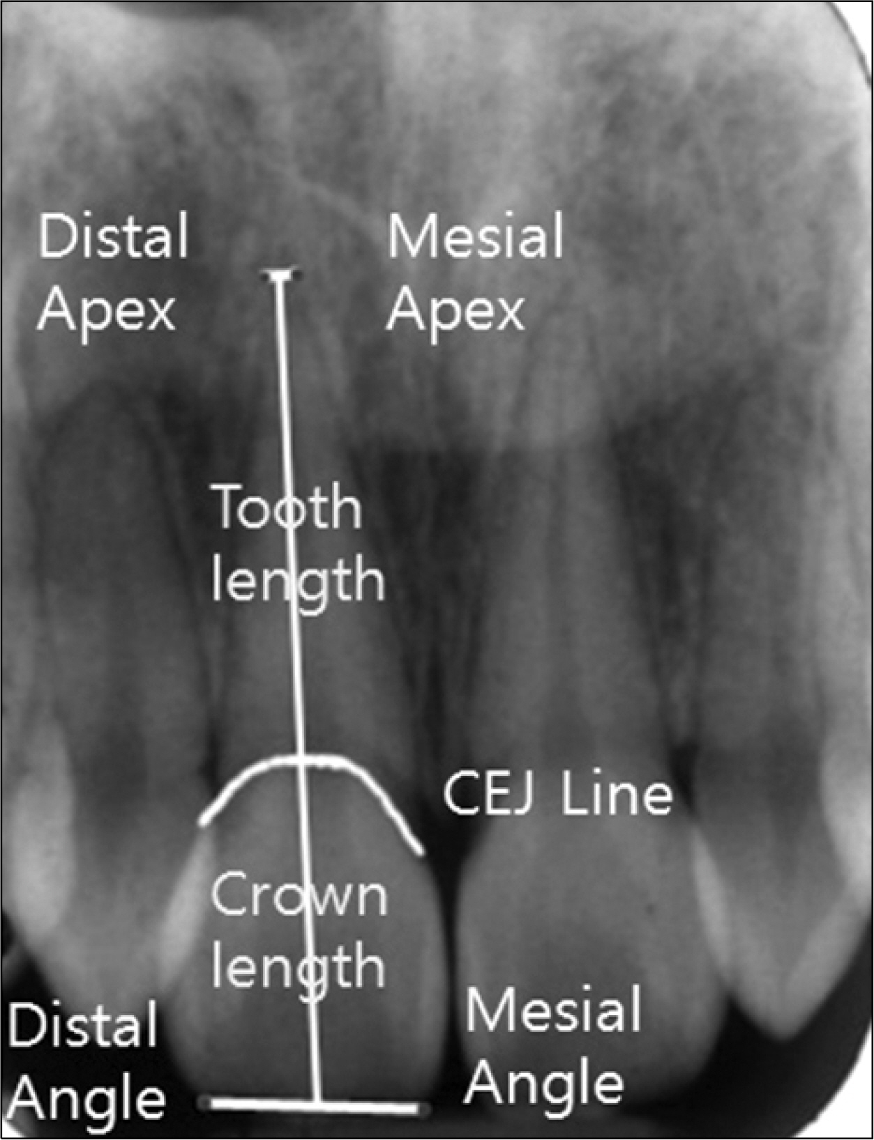
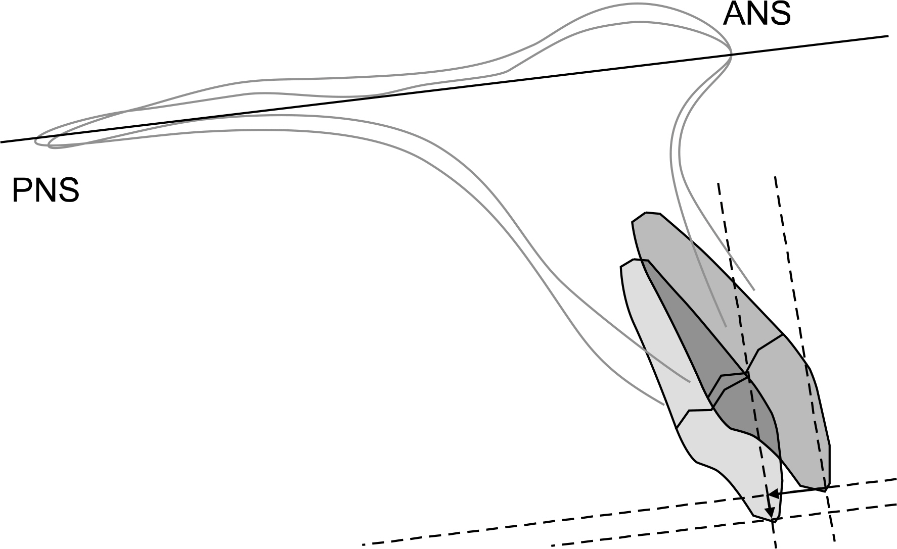
The records of 137 patients treated with full, fixed edgewise appliances were obtained from the Department of Orthodontics, Dankook University Dental Hospital, from November 2007 to December 2008. Periapical radiographs of the maxillary central incisors and cephalometric radiographs of each patient were used to assess apical root resorption and type of tooth movement.
RESULTS
The mean amount of resorption was 1.62 +/- 1.58 mm. The amount of resorption in the extraction and non-extraction groups was 2.10 +/- 1.64 mm and 1.18 +/- 1.39 mm, respectively. The amount of root resorption increased with the total tooth length. Severe root resorption (> 4 mm) was related to abnormal root shape (blunt, pointed, or eroded).
CONCLUSIONS
The variables significantly related to OIRR were extraction, initial tooth length, and root shape.
Keyword
MeSH Terms
Figure
Cited by 1 articles
-
Cone-beam computed tomographic evaluation of mandibular incisor alveolar bone changes for the intrusion arch technique: A retrospective cohort research
Lin Lu, Jiaping Si, Zhikang Wang, Xiaoyan Chen
Korean J Orthod. 2024;54(2):79-88. doi: 10.4041/kjod23.173.
Reference
-
1.Pizzo G., Licata ME., Guiglia R., Giuliana G. Root resorption and orthodontic treatment. Review of the literature. Minerva Stomatol. 2007. 56(1-2):31–44.2.Ketcham AH. A progress report of an investigation of apical root resorption of vital permanent teeth. Int J Orthod. 1929. 15:310–28.
Article3.Kim SC. A study on the affecting factors on root resorption. Korean J Orthod. 1994. 24:649–58.4.Sameshima GT., Asgarifar KO. Assessment of root resorption and root shape: periapical vs panoramic films. Angle Orthod. 2001. 71:185–9.5.McFadden WM., Engstrom C., Engstrom H., Anholm JM. A study of the relationship between incisor intrusion and root shortening. Am J Orthod Dentofacial Orthop. 1989. 96:390–6.
Article6.Linge BO., Linge L. Apical root resorption in upper anterior teeth. Eur J Orthod. 1983. 5:173–83.
Article7.Baumrind S., Korn EL., Boyd RL. Apical root resorption in or-thodontically treated adults. Am J Orthod Dentofacial Orthop. 1996. 110:311–20.
Article8.Mirabella AD., Artun J. Risk factors for apical root resorption of maxillary anterior teeth in adult orthodontic patients. Am J Orthod Dentofacial Orthop. 1995. 108:48–55.
Article9.Sameshima GT., Sinclair PM. Predicting and preventing root resorption: Part II. Treatment factors. Am J Orthod Dentofacial Orthop. 2001. 119:511–5.
Article10.Segal GR., Schiffman PH., Tuncay OC. Meta analysis of the treatment-related factors of external apical root resorption. Orthod Craniofac Res. 2004. 7:71–8.
Article11.Phillips JR. Apical root resorption under orthodontic therapy. Angle Orthod. 1955. 25:1–22.13.Sameshima GT., Sinclair PM. Predicting and preventing root resorption: Part I. Diagnostic factors. Am J Orthod Dentofacial Orthop. 2001. 119:505–10.
Article14.Hendrix I., Carels C., Kuijpers-Jagtman AM., Van 'T Hof M. A radiographic study of posterior apical root resorption in orthodontic patients. Am J Orthod Dentofacial Orthop. 1994. 105:345–9.
Article15.Harris EF., Kineret SE., Tolley EA. A heritable component for external apical root resorption in patients treated orthodon-tically. Am J Orthod Dentofacial Orthop. 1997. 111:301–9.
Article16.Kjaer I. Morphological characteristics of dentitions developing excessive root resorption during orthodontic treatment. Eur J Orthod. 1995. 17:25–34.
Article17.Owman-Moll P., Kurol J., Lundgren D. Repair of orthodonti-cally induced root resorption in adolescents. Angle Orthod. 1995. 65:403–8.18.Mirabella AD., Artun J. Prevalence and severity of apical root resorption of maxillary anterior teeth in adult orthodontic patients. Eur J Orthod. 1995. 17:93–9.
Article19.Hartsfield JK Jr., Everett ET., Al-Qawasmi RA. Genetic factors in external apical root resorption and orthodontic treatment. Crit Rev Oral Biol Med. 2004. 15:115–22.
Article20.Ioannidou-Marathiotou I., Papadopoulos MA., Kondylidou-Sidira A., Kokkas A., Karagiannis V. Digital subtraction radiography of panoramic radiographs to evaluate maxillary central incisor root resorption after orthodontic treatment. World J Orthod. 2010. 11:142–52.21.Witcher TP., Brand S., Gwilliam JR., McDonald F. Assessment of the anterior maxillain orthodontic patients using upper anterior occlusal radiographs and dental panoramic tomography: a comparison. Oral Surg Oral Med Oral Pathol Oral Radiol Endod. 2010. 109:765–74.22.Taylor NG., Jones AG. Are anterior occlusal radiographs indicated to supplement panoramic radiography during an orthodontic assessment? Br Dent J. 1995. 179:377–81.
Article23.Gher ME., Richardson AC. The accuracy of dental radiographic techniques used for evaluation of implant fixture placement. Int J Periodontics Restorative Dent. 1995. 15:268–83.24.Hemley S. The incidence of root resorption of vital permanent teeth. J Dent Res. 1941. 20:133–41.
Article26.Brin I., Tulloch JF., Koroluk L., Philips C. External apical root resorption in Class II malocclusion: a retrospective review of 1-versus 2-phase treatment. Am J Orthod Dentofacial Orthop. 2003. 124:151–6.27.Brezniak N., Wasserstein A. Orthodontically induced inflammatory root resorption. Part II: The clinical aspects. Angle Orthod. 2002. 72:180–4.28.Parker RJ., Harris EF. Directions of orthodontic tooth movements associated with external apical root resorption of the maxillary central incisor. Am J Orthod Dentofacial Orthop. 1998. 114:677–83.
Article29.Faltin RM., Faltin K., Sander FG., Arana-Chavez VE. Ultrastructure of cementum and periodontal ligament after continuous intrusion in humans: a transmission electron microscopy study. Eur J Orthod. 2001. 23:35–49.
Article30.Dudic A., Giannopoulou C., Martinez M., Montet X., Kiliaridis S. Diagnostic accuracy of digitized periapical radiographs validated against micro-computed tomography scanning in evaluating orthodontically induced apical root resorption. Eur J Oral Sci. 2008. 116:467–72.
Article31.Chan E., Darendeliler MA. Physical properties of root cementum: Part 5. Volumetric analysis of root resorption craters after application of light and heavy orthodontic forces. Am J Orthod Dentofacial Orthop. 2005. 127:186–95.
Article32.Haney E., Gansky SA., Lee JS., Johnson E., Maki K., Miller AJ, et al. Comparative analysis of traditional radiographs and conebeam computed tomography volumetric images in the diagnosis and treatment planning of maxillary impacted canines. Am J Orthod Dentofacial Orthop. 2010. 137:590–7.
Article
- Full Text Links
- Actions
-
Cited
- CITED
-
- Close
- Share
- Similar articles
-
- Factors affecting external apical root resorption of maxillary incisors associated with microimplantassisted rapid palatal expansion
- A radiographic study on root resorption in the malocclusion patients before orthodontic treatment
- Retrospective Analysis of Incisor Root Resorption Associated with Impacted Maxillary Canines
- Unilateral maxillary central incisor root resorption after orthodontic treatment for Angle Class II, division 1 malocclusion with significant maxillary midline deviation: A possible correlation with root proximity to the incisive canal
- Changes of root lengths and crestal bone height in nail biting patients


