J Adv Prosthodont.
2011 Jun;3(2):106-109. 10.4047/jap.2011.3.2.106.
A simple technique to fabricate a surgical obturator restoring the defect in original anatomical form
- Affiliations
-
- 1Department of Prosthodontics, Government Dental College & Hospital, Nagpur, Maharashtra, India. shambharkar@gmail.com
- KMID: 1975086
- DOI: http://doi.org/10.4047/jap.2011.3.2.106
Abstract
- Oral cancer treatment involves the surgical removal of all or part of the maxilla, leaving the patient with a defect that compromises the integrity and function of the oral cavity. The postoperative restoration of esthetics, deglutition, and speech shortens recovery time in the hospital and expedites the patient's return to the community as a functioning member. The surgical obturator is the proven treatment option in such situations. This article describes a simple technique to fabricate a surgical obturator that restores patient's original dentition and facial and palatal tissue form. The obturator fabricated with this technique utilizes the vacuum formed index of patient's original tissue form and duplicated partly in heat and partly in auto polymerizing acrylic resin. Duplication of the original tissue form helps patient to minimize the immense physiological trauma immediately after the surgical resection. The obturator fabricated with this technique supports soft tissues after surgery and minimizes scar contracture and disfigurement, and thus may have a positive effect on the patients' psychology.
Keyword
MeSH Terms
Figure
-
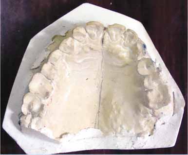
Fig. 1 Maxillary working cast with anticipated line of resection marked.
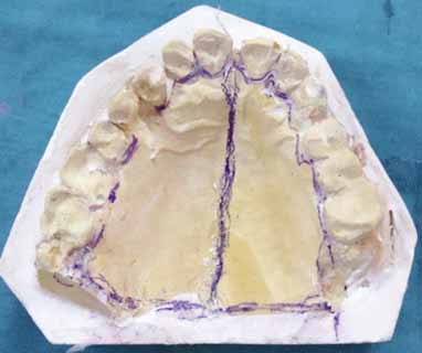
Fig. 2 Scraping of the cast to achieve the normal anatomical contours in the labial vestibule.
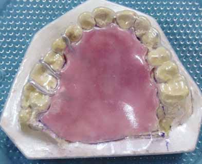
Fig. 3 Vacuum formed template adapted on the prosthetic base.
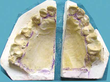
Fig. 4 Maxillary cast sectioned according to the pre-planned line of resection.
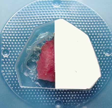
Fig. 5 Reseating of cast on vacuum formed template.
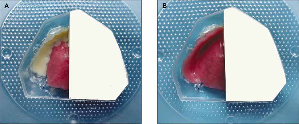
Fig. 6 Creation of the prosthetic teeth (A), and facial surface (B) in tooth colored and pink colored autopolymerizing acrylic resin respectively.
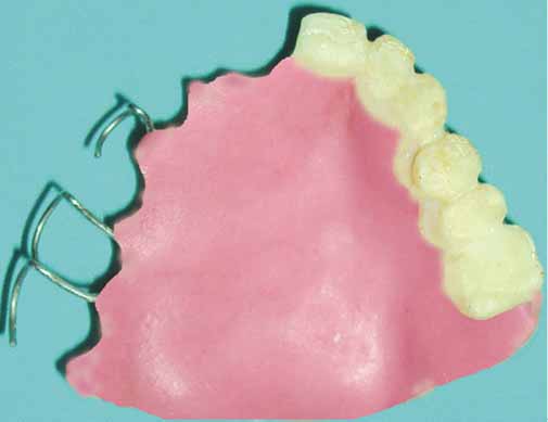
Fig. 7 Completed surgical obturator.
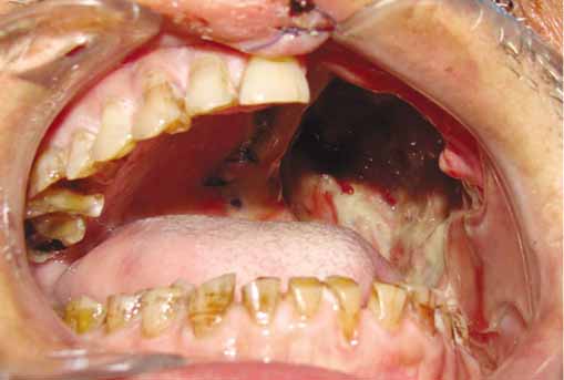
Fig. 8 Surgical defect after maxillectomy.

Fig. 9 Surgical obturator was placed after maxillectomy.
Reference
-
1. Kouyoumdjian JH, Chalian VA. An interim obturator prosthesis with duplicated teeth and palate. J Prosthet Dent. 1984. 52:560–562.2. DaBreo EL, Chalian VA, Lingeman R, Reisbick MH. Prosthetic and surgical management of osteogenic sarcoma of the maxilla. J Prosthet Dent. 1990. 63:316–320.3. Huryn JM, Piro JD. The maxillary immediate surgical obturator prosthesis. J Prosthet Dent. 1989. 61:343–347.4. King GE, Martin JW. Cast circumferential and wire clasps for obturator retention. J Prosthet Dent. 1983. 49:799–802.5. Wolfaardt JF. Modifying a surgical obturator prosthesis into an interim obturator prosthesis. A clinical report. J Prosthet Dent. 1989. 62:619–621.6. Arcuri MR, Taylor TD. Taylor TD, editor. Clinical management of the dentate maxillectomy patient. Clinical maxillofacial prosthetics. 2000. Carol Stream (IL): Quintessence;103–120.7. Beumer J, Curtis TA, Marunick MT. Maxillofacial rehabilitation: Prosthodontic and surgical considerations. 1996. St. Louis: Medico Dental Media International Inc..8. Gardner LK, Parr GR, Richardson DW. An interim buccal flange obturator. J Prosthet Dent. 1991. 65:862.9. Shaker KT. A simplified technique for construction of an interim obturator for a bilateral total maxillectomy defect. Int J Prosthodont. 2000. 13:166–168.10. Kaplan P. Stabilization of an interim obturator prosthesis using a denture duplicator. J Prosthet Dent. 1992. 67:377–379.11. DaBreo EL. A light-cured interim obturator prosthesis. A clinical report. J Prosthet Dent. 1990. 63:371–373.
- Full Text Links
-
- Actions
-
Cited
- CITED
-
- Close
- Share
-
- Similar articles
-
- Maxillofacial rehabilitation of hemi-maxillectomy patient using a closed hollow bulb obturator fabricated by one-step polymerization technique: a clinical report
- Use of artificial palate for improving facial support in the fabrication of a maxillary obturator: A case report
- Anatomical Reconstruction of the Medial Orbital Wall Fracture
- Strangulated Obturator Hernia: Mesh-Plug Technique
- Strangulated Obturator Hernia
