J Adv Prosthodont.
2011 Jun;3(2):69-75. 10.4047/jap.2011.3.2.69.
Comparative analysis of the clinical techniques used in evaluation of marginal accuracy of cast restoration using stereomicroscopy as gold standard
- Affiliations
-
- 1Department of Prosthodontics, Saraswati Dental College and Hospital, Lucknow, India. abhishek_rastogi82@yahoo.com
- KMID: 1975079
- DOI: http://doi.org/10.4047/jap.2011.3.2.69
Abstract
- PURPOSE
This study assessed the effect of preparation design on marginal adaptation and also compared the sensitivity and specificity of clinical evaluation techniques for marginal accuracy of cast restorations to stereomicroscopy.
MATERIALS AND METHODS
Three Ivorine molar teeth of different designs were prepared. (A)-A complete crown preparation with buccal shoulder and beveled finish line. (B)-A complete crown preparation with chamfer finish line. (C)-A three-quarter crown preparation with proximal boxes and beveled finish line. Twenty four castings were prepared with eight castings for each design respectively. Each casting underwent examination with an explorer, disclosing media, and a stereomicroscope. Stereomicroscopy at a value less than or equal to 30 microns was used as a gold standard to evaluate the significance of different designs on marginal adaptation. Chi-square tests of independence and Kruskal-Wallis were used to evaluate the effect of preparation design and compare the agreement between examination methods for detection of marginal gap size of greater than or equal to 30 microns (alpha=.05). Sensitivity and specificity for explorer and disclosing media as compared to stereomicroscope was calculated using statistical formula given by Park.
RESULTS
The preparation design did not significantly affect overall marginal adaptation. Examination by explorer and disclosing media at 30 microm revealed 39% and 10.06% sensitivity and 91% and 82% specificity respectively.
CONCLUSION
Preparation designs examined in this study did not significantly affect the marginal adaptation of the castings. Commonly used clinical evaluation techniques using explorer and disclosing media appeared to be inadequate for assessment of marginal accuracy.
Keyword
Figure
-
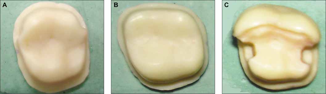
Fig. 1 Ivorine tooth preparations. Design A: complete crown preparation with a buccal shoulder and beveled finish line, Design B: complete crown preparation with a chamfer finish line, Design C: three-quarter crown preparation with proximal boxes and beveled finish line.

Fig. 2 Fit check using explorer.
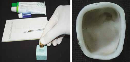
Fig. 3 Fit check using elastomeric disclosing media.

Fig. 4 Marginal gap as seen under stereomicroscope.

Fig. 5 Comparison of mean marginal openings of three designs in stereomicroscope.

Fig. 6 Percentage of fit and misfit sites in explorer, elastomeric disclosing media, and stereomicroscope.
Reference
-
1. Felton DA, Kanoy BE, Bayne SC, Wirthman GP. Effect of in vivo crown margin discrepancies on periodontal health. J Prosthet Dent. 1991. 65:357–364.2. Richter WA, Ueno H. Relationship of crown margin placement to gingival inflammation. J Prosthet Dent. 1973. 30:156–161.3. Preston JD. Rational approach to tooth preparation for ceramo-metal restorations. Dent Clin North Am. 1977. 21:683–698.4. Shillingburg HT Jr, Hobo S, Fisher DW. Preparation design and margin distortion in porcelain-fused-to-metal restorations. J Prosthet Dent. 1973. 29:276–284.5. Rosner D. Function, placement, and reproduction of bevels for gold castings. J Prosthet Dent. 1963. 13:1160–1166.6. Syu JZ, Byrne G, Laub LW, Land MF. Influence of finish-line geometry on the fit of crowns. Int J Prosthodont. 1993. 6:25–30.7. Rissin L, Wetreich G. Utilization of elastomeric materials to evaluate the accuracy of cast restorations prior to cementation. J Prosthet Dent. 1983. 49:585–586.8. Davis SH, Kelly JR, Campbell SD. Use of an elastomeric material to improve the occlusal seat and marginal seal of cast restorations. J Prosthet Dent. 1989. 62:288–291.9. White SN, Sorensen JA, Kang SK. Improved marginal seating of cast restorations using a silicone disclosing medium. Int J Prosthodont. 1991. 4:323–326.10. Walker PM. Remounting multiple casting prior to final cementation. J Prosthet Dent. 1981. 46:145–148.11. McLean JW, von Fraunhofer JA. The estimation of cement film thickness by an in vivo technique. Br Dent J. 1971. 131:107–111.12. Arakelian A Jr. A technique for seating castings. J Prosthet Dent. 1982. 48:357.13. Eames WB, O'Neal SJ, Monteiro J, Miller C, Roan JD Jr, Cohen KS. Techniques to improve the seating of castings. J Am Dent Assoc. 1978. 96:432–437.14. Belser UC, MacEntee MI, Richter WA. Fit of three porcelain-fused-to-metal marginal designs in vivo: a scanning electron microscope study. J Prosthet Dent. 1985. 53:24–29.15. Campagni WV, Wright W, Martinoff JT. Effect of die spacer on the seating of complete cast gold crowns with grooves. J Prosthet Dent. 1986. 55:324–328.16. Fusayama T. Technical procedure of precision casting. J Prosthet Dent. 1959. 9:1037–1048.17. Pascoe DF. Analysis of the geometry of finishing lines for full crown restorations. J Prosthet Dent. 1978. 40:157–162.18. Christensen GJ. Marginal fit of gold inlay castings. J Prosthet Dent. 1966. 16:297–305.19. Jahangiri L, Wahlers C, Hittelman E, Matheson P. Assessment of sensitivity and specificity of clinical evaluation of cast restoration marginal accuracy compared to stereomicroscopy. J Prosthet Dent. 2005. 93:138–142.20. Park K. Textbook of preventive and social medicine. 2005. 18th ed. Jabalpur: Banarsidas Bhanot publishers;117.
- Full Text Links
-
- Actions
-
Cited
- CITED
-
- Close
- Share
-
- Similar articles
-
- Comparison of the fit of cast gold crowns fabricated from the digital and the conventional impression techniques
- Three-dimensional accuracy of different correction methods for cast implant bars
- Estimation of relation between techniques of dye penetration for microleakage and SEM evaluation for marginal adaptation of the restoration
- A comparative study of gold UCLA-type and CAD/CAM titanium implant abutments
- THE EFFECTS OF FABRICATION AND TIGHTENING METHODS OF GOLD CYLINDER ON THE PRELOAD OF THE IMPLANT ABUTMENT
