Imaging Sci Dent.
2014 Dec;44(4):293-299. 10.5624/isd.2014.44.4.293.
Relationship between pain and effusion on magnetic resonance imaging in temporomandibular disorder patients
- Affiliations
-
- 1Department of Oral and Maxillofacial Radiology, School of Dentistry and Institute of Oral Bioscience, Chonbuk National University, Jeonju, Korea. kkj1512@jbnu.ac.kr
- KMID: 1974498
- DOI: http://doi.org/10.5624/isd.2014.44.4.293
Abstract
- PURPOSE
This study was performed to find the relationship between pain and joint effusion using magnetic resonance imaging (MRI) in temporomandibular disorder (TMD) patients.
MATERIALS AND METHODS
The study subjects included 232 TMD patients. The inclusion criteria in this study were the presence of spontaneous pain or provoked pain on one or both temporomandibular joints (TMJs). The provoked pain was divided into three groups: pain on palpation (G1), pain on mouth opening (G2), and pain on mastication (G3). MRI examinations were performed using a 1.5-T MRI scanner. T1- and T2-weighted images with para-sagittal and para-coronal images were obtained. According to the T2-weighted image findings, the cases of effusions were divided into four groups: normal, mild (E1), moderate (E2), and marked effusion (E3). A statistical analysis was carried out using the chi2 test with SPSS (version 12.0, SPSS Inc., Chicago, IL, USA).
RESULTS
Spontaneous pain, provoked pain, and both spontaneous and provoked pain were significantly related to joint effusion in TMD patients (p<0.05). However, among the various types of provoked pain, pain on palpation of the masticatory muscles and TMJ (G1) was not related to joint effusion in TMD patients (p>0.05).
CONCLUSION
Spontaneous pain was related to the MRI findings of joint effusion; however, among the various types of provoked pain, pain on palpation of the masticatory muscles and TMJ was not related to the MRI findings of joint effusion. These results suggest that joint effusion has a significant influence on the prediction of TMJ pain.
MeSH Terms
Figure
-
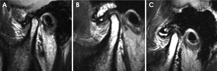
Fig. 1 T2-weighted magnetic resonance images show the joint effusion in the joint spaces. A. Mild effusion: a dot of high signal intensity in the superior joint space. B. Moderate effusion: between mild and marked effusion. C. Marked effusion: collection with pooling in the superior joint space.
Reference
-
1. Emshoff R, Rudisch A. Validity of clinical diagnostic criteria for temporomandibular disorders: clinical versus magnetic resonance imaging diagnosis of temporomandibular joint internal derangement and osteoarthrosis. Oral Surg Oral Med Oral Pathol Oral Radiol Endod. 2001; 91:50–55.2. Emshoff R, Brandlmaier I, Bertram S, Rudisch A. Relative odds of temporomandibular joint pain as a function of magnetic resonance imaging findings of internal derangement, osteoarthrosis, effusion, and bone marrow edema. Oral Surg Oral Med Oral Pathol Oral Radiol Endod. 2003; 95:437–445.
Article3. Bertram S, Rudisch A, Innerhofer K, Pümpel E, Grubwieser G, Emshoff R. Diagnosing TMJ internal derangement and osteoarthritis with magnetic resonance imaging. J Am Dent Assoc. 2001; 132:753–761.
Article4. Drace JE, Enzmann DR. Defining the normal temporomandibular joint: closed-, partially open-, and open-mouth MR imaging of asymptomatic subjects. Radiology. 1990; 177:67–71.
Article5. Haley DP, Schiffman EL, Lindgren BR, Anderson Q, Andreasen K. The relationship between clinical and MRI findings in patients with unilateral temporomandibular joint pain. J Am Dent Assoc. 2001; 132:476–481.
Article6. Rudisch A, Innerhofer K, Bertram S, Emshoff R. Magnetic resonance imaging findings of internal derangement and effusion in patients with unilateral temporomandibular joint pain. Oral Surg Oral Med Oral Pathol Oral Radiol Endod. 2001; 92:566–571.
Article7. Emshoff R, Brandlmaier I, Bertram S, Rudisch A. Risk factors for temporomandibular joint pain in patients with disc displacement without reduction - a magnetic resonance imaging study. J Oral Rehabil. 2003; 30:537–543.
Article8. Yano K, Sano T, Okano T. A longitudinal study of magnetic resonance (MR) evidence of temporomandibular joint (TMJ) fluid in patients with TMJ disorders. Cranio. 2004; 22:64–71.
Article9. Larheim TA. Role of magnetic resonance imaging in the clinical diagnosis of the temporomandibular joint. Cells Tissues Organs. 2005; 180:6–21.
Article10. Emshoff R, Rudisch A. Temporomandibular joint internal derangement and osteoarthrosis: are effusion and bone marrow edema prognostic indicators for arthrocentesis and hydraulic distention? J Oral Maxillofac Surg. 2007; 65:66–73.
Article11. Güler N, Yatmaz PI, Ataoglu H, Emlik D, Uckan S. Temporomandibular internal derangement: correlation of MRI findings with clinical symptoms of pain and joint sounds in patients with bruxing behaviour. Dentomaxillofac Radiol. 2003; 32:304–310.
Article12. Murakami K, Segami N, Fujimura K, Iizuka T. Correlation between pain and synovitis in patients with internal derangement of the temporomandibular joint. J Oral Maxillofac Surg. 1991; 49:1159–1162.
Article13. Takahashi T, Nagai H, Seki H, Fukuda M. Relationship between joint effusion, joint pain, and protein levels in joint lavage fluid of patients with internal derangement and osteoarthritis of the temporomandibular joint. J Oral Maxillofac Surg. 1999; 57:1187–1194.
Article14. Ohlmann B, Rammelsberg P, Henschel V, Kress B, Gabbert O, Schmitter M. Prediction of TMJ arthralgia according to clinical diagnosis and MRI findings. Int J Prosthodont. 2006; 19:333–338.15. Adame CG, Monje F, Offnoz M, Martin-Granizo R. Effusion in magnetic resonance imaging of the temporomandibular joint: a study of 123 joints. J Oral Maxillofac Surg. 1998; 56:314–318.
Article16. Westesson PL, Brooks SL. Temporomandibular joint: relationship between MR evidence of effusion and the presence of pain and disk displacement. AJR Am J Roentgenol. 1992; 159:559–559.
Article17. Suenaga S, Abeyama K, Hamasaki A, Mimura T, Noikura T. Temporomandibular disorders: relationship between joint pain and effusion and nitric oxide concentration in the joint fluid. Dentomaxillofac Radiol. 2001; 30:214–218.
Article18. Smith HJ, Larheim TA, Aspestrand F. Rheumatic and nonrheumatic disease in the temporomandibular joint: gadolinium-enhanced MR imaging. Radiology. 1992; 185:229–234.
Article19. Segami N, Suzuki T, Sato J, Miyamaru M, Nishimura M, Yoshimura H. Does joint effusion on T2 magnetic resonance images reflect synovitis? Part 3. Comparison of histologic findings of arthroscopically obtained synovium in internal derangements of the temporomandibular joint. Oral Surg Oral Med Oral Pathol Oral Radiol Endod. 2003; 95:761–766.
Article20. Koh KJ, List T, Petersson A, Rohlin M. Relationship between clinical and magnetic resonance imaging diagnoses and findings in degenerative and inflammatory temporomandibular joint diseases: a systemic literature review. J Orofac Pain. 2009; 23:123–139.21. Barclay P, Hollender LG, Maravilla KR, Truelove EL. Comparison of clinical and magnetic resonance imaging diagnosis in patients with disk displacement in the temporomandibular joint. Oral Surg Oral Med Oral Pathol Oral Radiol Endod. 1999; 88:37–43.22. Huddleston Slater JJ, Lobbezoo F, Chen YJ, Naeije M. A comparative study between clinical and instrumental methods for the recognition of internal derangements with a clicking sound on condylar movement. J Orofac Pain. 2004; 18:138–147.23. Uşüsumez S, Oz F, Güray E. Comparison of clinical and magnetic resonance imaging diagnoses in patients with TMD history. J Oral Rehabil. 2004; 31:52–56.24. Limchaichana N, Nilsson H, Ekberg EC, Nilner M, Petersson A. Clinical diagnoses and MRI findings in patients with TMD pain. J Oral Rehabil. 2007; 34:237–245.
Article25. Sano T, Westesson PL. Magnetic resonance imaging of the temporomandibular joint. Increased T2 signal in the retrodiskal tissue of painful joints. Oral Surg Oral Med Oral Pathol Oral Radiol Endod. 1995; 79:511–516.26. Harms SE, Wilk RM, Wolford LM, Chiles DG, Milam SB. The temporomandibular joint: magnetic resonance imaging using surface coils. Radiology. 1985; 157:133–136.
Article27. Emshoff R, Brandimaier I, Bertram S, Rudisch A. Magnetic resonance imaging findings of osteoarthrosis and effusion in patients with unilateral temporomandibular joint pain. Int J Oral Maxillofac Surg. 2002; 31:598–602.
Article28. Emshoff R, Puffer P, Rudisch A, Gassner R. temporomandibular joint pain: relationship to internal derangement type, osteoarthrosis, and synovial fluid mediator level of tumor necrosis factor-alpha. Oral Surg Oral Med Oral Pathol Oral Radiol Endod. 2000; 90:442–449.29. Güler N, Uçkan S, Imirzalioğlu P, Açikgözoğlu S. Temporomandibular joint internal derangement: relationship between joint pain and MR grading of effusion and total protein concentration in the joint fluid. Dentomaxillofac Radiol. 2005; 34:175–181.
Article30. Nishimura M, Segami N, Kaneyama K, Suzuki T, Miyamaru M. Relationships between pain-related mediators and both synovitis and joint pain in patients with internal derangements and osteoarthritis of the temporomandibular joint. Oral Surg Oral Med Oral Pathol Oral Radiol Endod. 2002; 94:328–332.
Article
- Full Text Links
-
- Actions
-
Cited
- CITED
-
- Close
- Share
-
- Similar articles
-
- Magnetic resonance evidence of joint effusion in patients with temporomandibular joint disorders
- Analysis of the clinical symptoms and the temporomandibular joint disk by magnetic resonance imaging after conservative treatment with anterior repositioning splint
- Comparison of MRI findings with clinical symptoms in temporomandibular joint internal derangement
- Relationship between anterior disc displacement with/without reduction and effusion in temporomandibular disorder patients using magnetic resonance imaging
- Effusion in magnetic resonance imaging of the temporomandibular joint
