Osteosarcoma of the mandible: A case report with an early radiographic manifestation
- Affiliations
-
- 1Department of Oral Pathology and Microbiology, Mahatma Gandhi Post Graduate Institute of Dental Sciences, Pondicherry, India.
- 2Department of Oral and Maxillofacial Pathology, Chettinad Dental College and Research Institute, Tamilnadu, India. ksrirammds@gmail.com
- KMID: 1974479
- DOI: http://doi.org/10.5624/isd.2014.44.1.85
Abstract
- Osteosarcoma is a classical malignant bone-forming neoplasm which usually presents with an aggressive clinical course. The current case is presented with the radiographic feature of widening of the periodontal ligament space of the involved teeth, which is considered to be the earliest radiographic manifestation of osteosarcoma involving the jaw bone. The main aim of this case report was to focus on the importance of early diagnosis of this tumor based on clinical and radiographic examinations, and confirmation by histopathology. Considering the rarity of the disease type and particularly taking into account the fast progression and aggressiveness of this neoplasm, it is clear that the presentation of a clinical case represents a major contribution to better understanding of osteosarcomas involving the jaw bone.
Keyword
Figure
-

Fig. 1 A photograph shows a soft tissue growth on the right body of the mandible.

Fig. 2 A panoramic radiograph shows the loss of the lamina dura and widening of the periodontal ligament space involving the right permanent mandibular first molar and second premolar with mild rarefaction of the interdental alveolar bone between the right mandibular first molar and second molar.

Fig. 3 A periapical radiograph shows the loss of the lamina dura and periradicular bone loss involving the interdental bone between the right permanent mandibular second premolar and first molar.
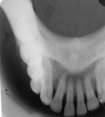
Fig. 4 A right mandibular lateral occlusal radiograph shows the shadow of the soft tissue mass on the right side and peripheral cortical reactive bone formation.

Fig. 5 A. A photomicrograph shows neoplastic osteiod formation with varying degrees of calcification (H&E stain, 10×). B. A photomicrograph shows pleomorphic and hyperchromatic malignant osteoblastic cells (H&E stain, 40×).
Reference
-
1. Khorate MM, Goel S, Singh MP, Ahmed J. Osteosarcoma of mandible: a case report and review of literature. J Cancer Sci Ther. 2010; 2:122–125.2. Ong ST, Shim CK, Ng KH, Siar CH. Osteosarcoma presenting as an aggressive nodular mass in the region of the mandible. J Oral Sci. 2004; 46:55–59.
Article3. Neville BW, Damm DD, Allen CM, Bouquot JE. Oral and maxillofacial pathology. 3rd ed. St. Louis: Saunders/Elsevier;2009. p. 517–519.4. Donaldson ME, Geist JR, Daley TD. Osteosarcoma of the jaws in children. Int J Paediatr Dent. 2004; 14:54–60.
Article5. Bras JM, Donner R, van der Kwast WA, Snow GB, van der Waal I. Juxtacortical osteogenic sarcoma of the jaws. Review of the literature and report of a case. Oral Surg Oral Med Oral Pathol. 1980; 50:535–544.6. Anil S, Krishnan AP, Rajendran R. Osteosarcoma of the mandible masquerading as a dental abscess: report of a case. Case Rep Dent. 2012; 2012:635062.
Article7. Amaral MB, Buchholz I, Freire-Maia B, Reher P, de Souza PE, Marigo Hde A, et al. Advanced osteosarcoma of the maxilla: a case report. Med Oral Patol Oral Cir Bucal. 2008; 13:E492–E495.8. Marx RE, Stern D. Oral and Maxillofacial Pathology: a rationale for diagnosis and treatment. Chicago: Quintessence Pub Co;2003. p. 805–806.
