Gold-wire artifacts on diagnostic radiographs: A case report
- Affiliations
-
- 1Department of Periodontology, Department of Oral Health Sciences, KU Leuven and Dentistry, University Hospitals, KU Leuven, Kapucijnenvoer 7, 3000 Leuven, Belgium. marc.quirynen@med.kuleuven.be
- 2Oral Imaging Centre, Department of Oral Health Sciences, KU Leuven and Dentistry, University Hospitals, KU Leuven, Kapucijnenvoer 7, 3000 Leuven, Belgium.
- KMID: 1974478
- DOI: http://doi.org/10.5624/isd.2014.44.1.81
Abstract
- This report described a case in which diagnostic radiographs showed irregular dense radiopaque strings and curved lines in the head and neck area. These artifacts could lead to misinterpretation since they may obscure anatomical structures and/or mask critical structures/pathologies. A more detailed history of the patient indicated that these strings originated from a facelift procedure in which a gold-wire technique was used. Considering that such intervention may cause a radiodiagnostic burden, it should be included in the anamnesis prior to radiography.
Keyword
Figure
-

Fig. 1 Photographs show a 76-year-old patient who presented with normal facial skin, with only few wrinkles on the cheek.

Fig. 2 Intraoral radiographs of the posterior part of the first quadrant show irregular strings and curved lines of a dense radiopaque material.
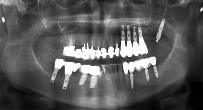
Fig. 3 A panoramic image shows irregular dense radiopaque strings on the left and the right side. The periodontal and peri-implant tissues seem healthy.

Fig. 4 A. A coronal cone-beam computed tomography image, on the position of the second premolar, clearly highlights the radiopaque structures in the left and the right cheek, lateral to the buccinator and masseter muscles. B. A three-dimensional reconstruction image clearly visualizes the gold wires, besides the artifacts caused by the implants and partial bridges.
Reference
-
1. Murray D, Whyte A. Dental panoramic tomography: what the general radiologist needs to know. Clin Radiol. 2002; 57:1–7.
Article2. Armstrong DT, Gilliland WR, Murphey MD, Salesky J. Clinical images: gold thread acupuncture for rheumatoid arthritis. Arthritis Rheum. 2011; 63:3021.
Article3. Brown RS, Coleman-Bennett MM, Jones-Matthews T. Synthetic hair braid extension artifacts in panoramic radiographs. J Am Dent Assoc. 1998; 129:601–604.
Article4. Scheifele C, Lemke AJ, Reichart PA. Hair artefacts in the head and neck region. Dentomaxillofac Radiol. 2003; 32:255–257.
Article5. Myllylä RM, Haapasaari KM, Palatsi R, Germain-Lee EL, Hägg PM, Ignatius J, et al. Multiple miliary osteoma cutis is a distinct disease entity: four case reports and review of the literature. Br J Dermatol. 2011; 164:544–552.
Article6. Gfesser M, Worret WI, Hein R, Ring J. Multiple primary osteoma cutis. Arch Dermatol. 1998; 134:641–643.7. Goldminz D, Greenberg RD. Multiple miliary osteoma cutis. J Am Acad Dermatol. 1991; 24:878–881.
Article8. de Moura MD, Madureira DF, Noman-Ferreira LC, Abdo EN, de Aguiar EG, Freire AR. Tonsillolith: a report of three clinical cases. Med Oral Patol Oral Cir Bucal. 2007; 12:E130–E133.9. Alsaadi G, Jacobs R, Quirynen M, van Steenberghe D. Soft tissue augmentation of the cheeks detected on intra- and extraoral radiographs: a case report. Dentomaxillofac Radiol. 2008; 37:117–120.
Article10. Valiyaparambil J, Rengasamy K, Mallya SM. An unusual soft tissue radiopacity - radiographic appearance of a dermal filler. Br Dent J. 2009; 207:211–212.
Article11. Mupparapu M, Mozaffari E. Bilateral calcifications secondary to synthetic soft tissue augmentation of the cheeks: report of a case. Dentomaxillofac Radiol. 2002; 31:388–390.
Article12. Altuğ HA, Büyüksoy V, Okçu KM, Doğan N. Hemangiomas of the head and neck with phleboliths: clinical features, diagnostic imaging, and treatment of 3 cases. Oral Surg Oral Med Oral Pathol Oral Radiol Endod. 2007; 103:e60–e64.
Article13. Ikegami N, Nishijima K. Hemangioma of the buccal pad with phlebolithiasis: report of a case. Acta Med Okayama. 1984; 38:79–87.14. Smith JF, Drake J, Sollee N. Massive oral hemangioma with phlebolithiasis. Oral Surg Oral Med Oral Pathol. 1966; 21:83–88.
Article15. Klein AW. Soft tissue augmentation 2006: filler fantasy. Dermatol Ther. 2006; 19:129–133.
Article16. Newman J. Review of soft tissue augmentation in the face. Clin Cosmet Investig Dermatol. 2009; 2:141–150.
Article17. Coleman SR. Structural fat grafts: the ideal filler? Clin Plast Surg. 2001; 28:111–119.18. Carruthers J, Cohen SR, Joseph JH, Narins RS, Rubin M. The science and art of dermal fillers for soft-tissue augmentation. J Drugs Dermatol. 2009; 8:335–350.19. Burgess CM. Principles of soft tissue augmentation for the aging face. Clin Interv Aging. 2006; 1:349–355.
Article20. goldthread.ru [Internet]. Cincinnati: Gold Thread Worldwide;c2007-2014. cited 2012 August 5. Available from: http://www.goldthread.ru/eng.
- Full Text Links
-
- Actions
-
Cited
- CITED
-
- Close
- Share
-
- Similar articles
-
- Determination and classification of intraoral phosphor storage plate artifacts and errors
- External Fixator and External Fixator Supplemented with K-wire in the Treatment of Distal Radius Fractures
- Removal of a Broken Intramedullary Nail with a Narrow Hollow Using a Bulb-tipped Guide Wire and Kirschner Wire: A Case Report
- An Unusual Case of Guide Wire Fracture during Coronary Artery Stenting for Bifurcation Lesion
- Migration of a Broken Wire to the Popliteal Fossa After the Osteosynthesis of Patella Fracture: A Case Report
