Determination and classification of intraoral phosphor storage plate artifacts and errors
- Affiliations
-
- 1Department of Dental and Maxillofacial Radiology, Faculty of Dentistry, Çanakkale Onsekiz Mart University, Çanakkale, Turkey. yesimdeniz@comu.edu.tr
- 2Department of Restorative Dentistry, Faculty of Dentistry, Alanya Alaaddin Keykubat University, Antalya, Turkey.
- KMID: 2458371
- DOI: http://doi.org/10.5624/isd.2019.49.3.219
Abstract
- PURPOSE
The aim of this study was to determine the reasons and solutions for intraoral phosphor storage plate (PSP) image artifacts and errors, and to develop an appropriate classification of the artifacts.
MATERIALS AND METHODS
This study involved the retrospective examination of 5,000 intraoral images that had been obtained using a phosphor plate system. Image artifacts were examined on the radiographs and classified according to possible causative factors.
RESULTS
Artifacts were observed in 1,822 of the 5,000 images. After examination of the images, the errors were divided into 6 groups based on their causes, as follows: images with operator errors, superposition of undesirable structures, ambient light errors, plate artifacts (physical deformations and contamination), scanner artifacts, and software artifacts. The groups were then re-examined and divided into 45 subheadings.
CONCLUSION
Identification of image artifacts can help to improve the quality of the radiographic image and control the radiation dose. Knowledge of the basic physics and technology of PSP systems could aid to reduce the need for repeated radiography.
Keyword
MeSH Terms
Figure
-

Fig. 1 A. Phalangioma (arrow). B. The dental fillings (highest attenuation value areas) lost photons and had a shiny appearance when exposed to ambient light. C. A text pattern caused by exposure to visible light.

Fig. 2 Phosphor storage plate (PSP) damage. A. Stretches. B. Crescent-shaped bending (arrow). C. Bite marks (arrow). D. Partial stripping (arrow). E. Peeled plate surface. F. Reticulation caused by a peeled PSP surface. G. Elongation of the image resulting from use of a mismatched plate.
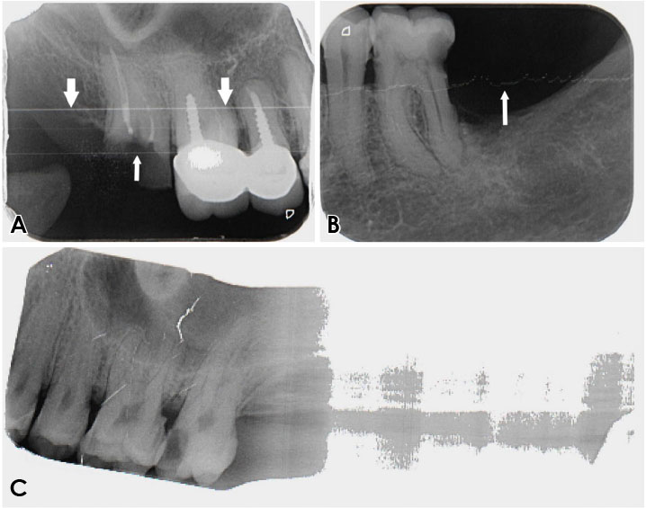
Fig. 3 Scanner artifacts. A. The gray lines extend in the fast scan direction (thick arrow) (caused by dust on the light guide) and dust particles (thin arrow). B. The gray line extends in the fast scan direction (arrow). C. Transport belt artifact (caused by the belt motor not moving at the correct speed, resulting in an abnormal elongation of the image).
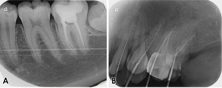
Fig. 4 Scanner artifacts. A. Laser slit dirt. B. Eraser unit artifact.

Fig. 5 Scanner artifacts. A–C. Laser unit fault.
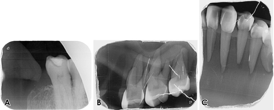
Fig. 6 Scanner artifacts. A. Straight radiolucent lines (with the likely cause being dust/dirt particles on the pickup light guide). B and C. Parallel multiple radiolucent lines images obtained from a phantom head. The likely cause was electromagnetic interference.

Fig. 7 Software artifacts. A. An edge masking defect causing a noisy view. B. An edge masking defect simulating a cone-cut image.
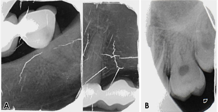
Fig. 8 Software artifacts. A. A bisection artifact (dividing the same image into two images). B. Plate size determination error. The reason for this artifact is that the scanner detected a different size from that of the scanned plate.
Reference
-
1. Wakoh M, Kuroyanagi K. Digital imaging modalities for dental practice. Bull Tokyo Dent Coll. 2001; 42:1–14.
Article2. Mol A. Digital Imaging. In : Mallya SM, Lam EW, editors. White and Pharoah's oral radiology: principles and interpretation. 8rd ed. St. Louis: Elsevier;2019. p. 40–60.3. Çalışkan A, Sumer AP. Definition, classification and retrospective analysis of photostimulable phosphor image artefacts and errors in intraoral dental radiography. Dentomaxillofac Radiol. 2017; 46:20160188.
Article4. Chiu HL, Lin SH, Chen CH, Wang WC, Chen JY, Chen YK, et al. Analysis of photostimulable phosphor plate image artifacts in an oral and maxillofacial radiology department. Oral Surg Oral Med Oral Pathol Oral Radiol Endod. 2008; 106:749–756.
Article5. Gulsahi A, Secgin CK. Assessment of intraoral image artifacts related to photostimulable phosphor plates in a dentomaxillofacial radiology department. Niger J Clin Pract. 2016; 19:248–253.
Article6. Shetty CM, Barthur A, Kambadakone A, Narayanan N, Kv R. Computed radiography image artifacts revisited. AJR Am J Roentgenol. 2011; 196:W37–W47.
Article7. Langland OE, Langlais RP, Preece JW. Analysis of errors and artifacts. Principles of dental imaging. Baltimore: Williams & Wilkins;2002. p. 160.
Article8. Kavo.com [Internet]. Biberach: SOREDEX™ DIGORA™ Optime;c2019. cited 2019 Jan 7. Available from: https://www.kavo.com/dental-x-ray-machines-and-diagnostics/scan-exam-intraoral#docs.9. Duerrdental.com [Internet]. Bietigheim-Bissingen: VistaScan Mini Easy;Available from: https://www.duerrdental.com/en/products/imaging/vistascan-image-plate-scanner/vistascan-mini-easy/.10. Seibert JA, Bogucki TM, Ciona T, Huda W, Karellas A, Mercier JR, et al. Report No. 093 - Acceptance testing and quality control of photostimulable storage phosphor imaging systems. Alexandria: American Association of Physicists in Medicine;2006.11. Seeram E. Computed radiography: physics and technology. Digital radiography. Singapore: Springer;2019. p. 41–63.12. Duckworth JE, Judy PF, Goodson JM, Socransky SS. A method for the geometric and densitometric standardization of intraoral radiographs. J Periodontol. 1983; 54:435–440.
Article13. Webber RL, Ruttimann UE, Groenhuis RA. Computer correction of projective distortions in dental radiographs. J Dent Res. 1984; 63:1032–1036.
Article14. Menchini-Fabris GB, Covani U, Toti P, Marconcini S, Barone A, Martuscelli R. A methodological approach to standardize and control the quality of the position and alignment of lamina implants on two-dimensional radiographs. Oral Radiol. (in press).
Article15. Sanghvi S, Rao A, Karuna YM, Shetty K, Nayak A. Image distortion of the roots of maxillary deciduous molars on Iopa's recorded with bisecting angle or a modified paralleling technique. Biomed Pharmacol J. 2018; 11:1039–1042.
Article16. Akdeniz BG, Gröndahl HG, Kose T. Effect of delayed scanning of storage phosphor plates. Oral Surg Oral Med Oral Pathol Oral Radiol Endod. 2005; 99:603–607.
Article17. Roberts MW, Mol A. Clinical techniques to reduce sensor plate damage in PSP digital radiography. J Dent Child (Chic). 2004; 71:169–170.18. Kalathingal SM, Shrout MK, Comer C, Brady C. Rating the extent of surface scratches on photostimulable storage phosphor plates in a dental school environment. Dentomaxillofac Radiol. 2010; 39:179–183.
Article
- Full Text Links
-
- Actions
-
Cited
- CITED
-
- Close
- Share
-
- Similar articles
-
- Artifacts in Grayscale Ultrasound
- Artifacts and Pitfalls in Neuromusculoskeletal Ultrasound
- Intraoral digital radiography: A comprehensive report on the technical specifications of current and historical systems
- Digital Chest Radiography with an Amorphous Silicon Flat-Panel-Detector Versus a Storage-Phosphor System: Comparison of Soft-Copy Images
- A Study on the Diagnostic Detection Ability of the Artificial Proximal Caries by Digora(R)
