Arthroscopic Treatment of Subchondral Bony Cyst in Early Osteoarthritis of the Hip Joint Using Allogeneic Bone Graft: A Report of Two Cases
- Affiliations
-
- 1Department of Orthopedic Surgery, Chungnam National University School of Medicine, Daejeon, Korea. dshwang@cnu.ac.kr
- 2Department of Orthopedic Surgery, Konyang University College of Medicine, Daejeon, Korea.
- KMID: 1974389
- DOI: http://doi.org/10.5371/hp.2015.27.2.110
Abstract
- Subchondral bony cyst, large solitary or multiple cysts in acetabular dome usually exacerbate progression to degenerative osteoarthritis in the hip joint. But it can be treated through arthroscopic intervention. We report two cases that treated by arthroscopic curettage and bone graft for subchondral bony cysts in early osteoarthritis of the hip joint, and it may delay progression to moderate osteoarthritis.
Keyword
Figure
-
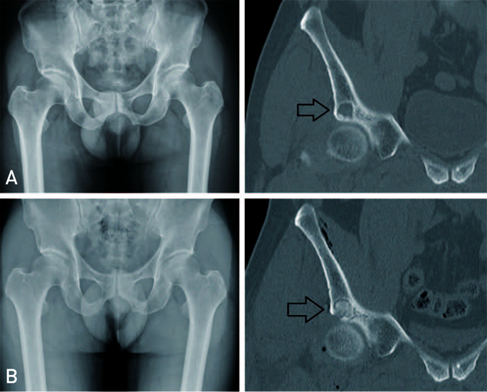
Fig. 1 (A) Preoperative simple X-ray and computed tomography (CT) image of Case 1. A large cyst (arrow) is located on the acetabular dome. (B) Postoperative immediately CT image and plain radiographs at last follow-up. The bone graft appears well maintained (arrow).
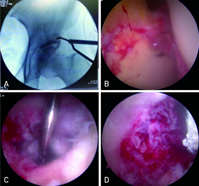
Fig. 2 Cyst location and arthroscopic procedure. (A) A C-arm image intensifier allows the cyst to be located. (B) Osteoarthritis finding in arthroscopy. (C) Opening of the cyst and the yellowish serous fluid drained and curettage of the outer and inner surface of the cyst. (D) Allogeneic bone graft inside the cyst using a silicon tube.
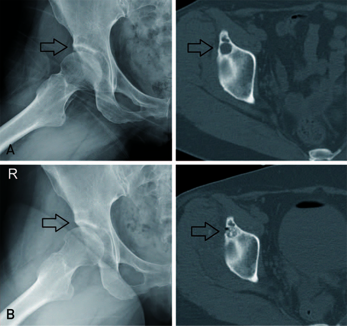
Fig. 3 (A) Preoperative simple X-ray and computed tomography (CT) image for Case 2. Multiple cysts (arrow) are revealed on the acetabular dome. (B) Postoperative immediately CT image and plain radiograph at last follow-up. The allogeneic bone graft is well maintained at its initial location (arrows).
Reference
-
1. Sabokbar A, Crawford R, Murray DW, Athanasou NA. Macrophage-osteoclast differentiation and bone resorption in osteoarthrotic subchondral acetabular cysts. Acta Orthop Scand. 2000; 71:255–261.
Article2. Ondrouch AS. Cyst formation in osteoarthritis. J Bone Joint Surg Br. 1963; 45:755–760.
Article3. Dürr HD, Martin H, Pellengahr C, Schlemmer M, Maier M, Jansson V. The cause of subchondral bone cysts in osteoarthrosis: a finite element analysis. Acta Orthop Scand. 2004; 75:554–558.
Article4. Leunig M, Beck M, Kalhor M, Kim YJ, Werlen S, Ganz R. Fibrocystic changes at anterosuperior femoral neck: prevalence in hips with femoroacetabular impingement. Radiology. 2005; 236:237–246.
Article5. Schmalzried TP, Akizuki KH, Fedenko AN, Mirra J. The role of access of joint fluid to bone in periarticular osteolysis. A report of four cases. J Bone Joint Surg Am. 1997; 79:447–452.6. Jamali AA, Fritz AT, Reddy D, Meehan JP. Minimally invasive bone grafting of cysts of the femoral head and acetabulum in femoroacetabular impingement: arthroscopic technique and case presentation. Arthroscopy. 2010; 26:279–285.
Article7. Ferguson AB Jr. The pathology of degenerative arthritis of the hip and the use of osteotomy in its treatment. Clin Orthop Relat Res. 1971; 77:84–97.8. Milgram JW. Morphologic alterations of the subchondral bone in advanced degenerative arthritis. Clin Orthop Relat Res. 1983; 173:293–312.9. Yoshida M, Konishi N. Subchondral cysts arise in the anterior acetabulum in dysplastic osteoarthritic hips. Clin Orthop Relat Res. 2002; 404:291–301.
Article10. McBeath AA, Neidhart DA. Acetabular cyst with communicating ganglion. A case report. J Bone Joint Surg Am. 1976; 58:267–269.
Article
- Full Text Links
-
- Actions
-
Cited
- CITED
-
- Close
- Share
-
- Similar articles
-
- A Large Subchondral Cyst in an Osteoarthritic Temporomandibular Joint: A Case Report
- Pathologic Fracture in a Subchondral Cystic Lesion at the Superior Acetabulum
- Arthroscopic Management for Septic Arthritis of the Hip Joint in Adults: A Report of Four Cases
- Unusual Cause of Hip Pain: Intrusion of the Acetabular Labrum
- Osteochondritis Dissecans of the Knee
