Analyses Using Micro-CT Scans and Tissue Staining on New Bone Formation and Bone Fusion According to the Timing of Cranioplasty via Frozen Autologous Bone Flaps in Rabbits : A Preliminary Report
- Affiliations
-
- 1Department of Neurosurgery, Kyung Hee University Hospital at Gangdong, Kyung Hee University School of Medicine, Seoul, Korea. toast2000@hanmail.net
- 2Department of Oral and Maxillofacial Surgery, Kyung Hee University Dental Hospital at Gangdong, Kyung Hee University School of Dentistry, Seoul, Korea.
- KMID: 1956421
- DOI: http://doi.org/10.3340/jkns.2015.57.4.242
Abstract
OBJECTIVE
The timing of cranioplasty and method of bone flap storage are known risk factors of non-union and resorption of bone flaps. In this animal experimental study, we evaluated the efficacy of cranioplasty using frozen autologous bone flap, and examined whether the timing of cranioplasty after craniectomy affects bone fusion and new bone formation.
METHODS
Total 8 rabbits (male, older than 16 weeks) were divided into two groups of early cranioplasty group (EG, 4 rabbits) and delayed cranioplasty group (DG, 4 rabbits). The rabbits of each group were performed cranioplasty via frozen autologous bone flaps 4 weeks (EG) and 8 weeks (DG) after craniectomy. In order to obtain control data, the cranioplasty immediate after craniectomy were made on the contralateral cranial bone of the rabbits (control group, CG).The bone fusion and new bone formation were evaluated by micro-CT scan and histological examination 8 weeks after cranioplasty on both groups.
RESULTS
In the micro-CT scans, the mean values of the volume and the surface of new bone were 50.13+/-7.18 mm3 and 706.23+/-77.26 mm2 in EG, 53.78+/-10.86 mm3 and 726.60+/-170.99 mm2 in DG, and 31.51+/-12.84 mm3 and 436.65+/-132.24 mm2 in CG. In the statistical results, significant differences were shown between EG and CG and between DG and CG (volume : p=0.028 and surface : p=0.008). The histological results confirmed new bone formation in all rabbits.
CONCLUSION
We observed new bone formation on all the frozen autologous bone flaps that was stored within 8 weeks. The timing of cranioplasty may showed no difference of degree of new bone formation. Not only the healing period after cranioplasty but the time interval from craniectomy to cranioplasty could affect the new bone formation.
Keyword
Figure
-
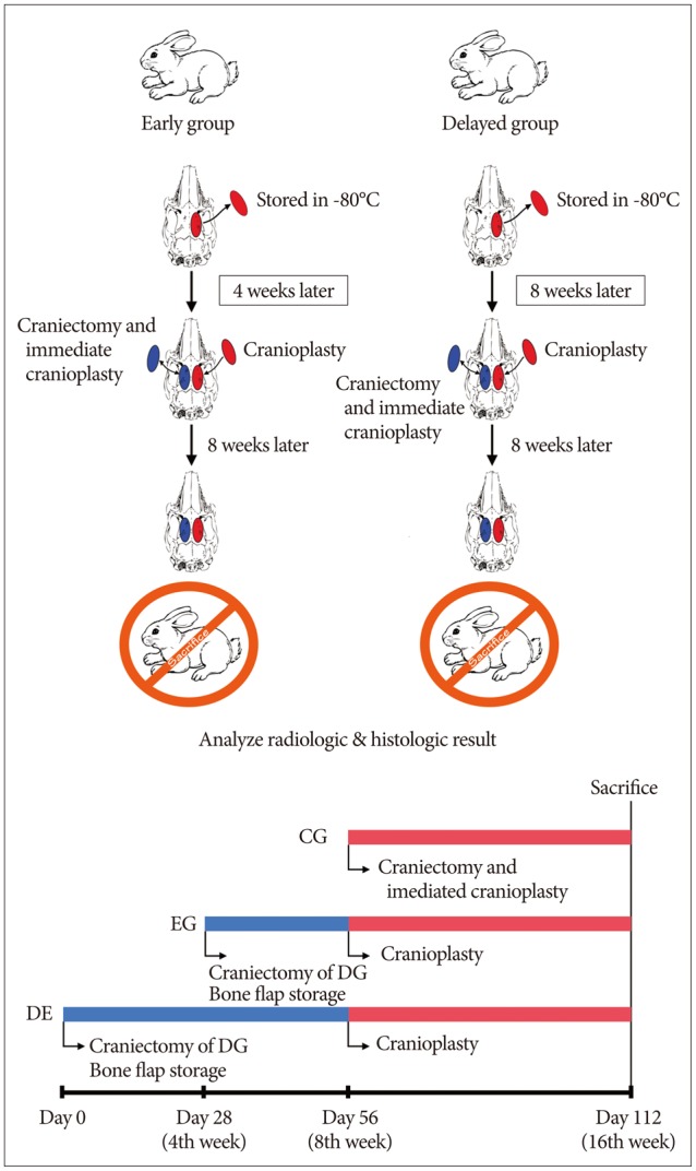
Fig. 1 Diagram and time-line of experimental schedule. Eight rabbits were divided into two groups of the delayed cranioplasty group (DG, 4 rabbits) and the early cranioplasty group (EG, 4 rabbits). The craniectomy was performed on the right side cranial bone of the DG rabbit at day 0, and EG rabbit at day 28. The obtained bone flap was stored in a freezer of -80℃. At day 56, cranioplasty was performed on both groups via the frozen bone flap. In order to obtain control data, the craniectomy was made on the left cranial bone of the rabbit, and the bone flap was immediately fixed (control group). At day 112, the EG and DG rabbits were sacrificed.
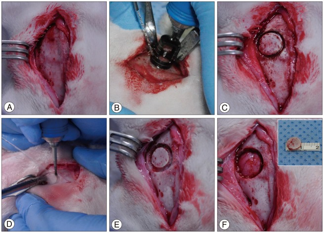
Fig. 2 Preparation of the experimental model of formation of bone defect and bone flap. After incision was made along the midline of the cranial skin, the muscles and periosteum were incised layer by layer to expose the cranial bone (A). The bone flap margin was formed 3-mm lateral to the sagittal suture, using a trephine drill with a 10-mm outer diameter and 9-mm inner diameter. To avoid damaging the dura mater, the inner table of the cortical bone was left (B and C). Using a 1-mm round burr, the space between the bone-flap margin and the cranial bone was expanded out of the bone flap in order to create a 12-mm bone defect diameter (D), and the dura mater was carefully exposed (E). The 9-mm bone flap was lifted up and the bone defect was formed (F).
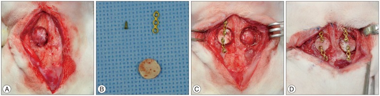
Fig. 3 The fibrous tissues were removed in order to expose the bone defect area (A). Afterwards, the autologous bone flap (B), which has been kept in the freezer, was applied to each labeled rabbit in order to perform cranioplasty, and the bone flap was fixed to cranial bone using a titanium-alloy miniplate and screws (C). Using the same method, the control group was formed on the left cranium of the rabbit (D).
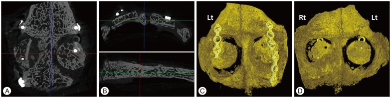
Fig. 4 The axial sectional image (A), and coronal and sagittal sectional images (B) of computed tomography scans. The 3D reconstructed images showed outer surface (C) and inner surface (D) of cranium. The left side of cranium is experimental craniectomy site, and the right side is the control group.
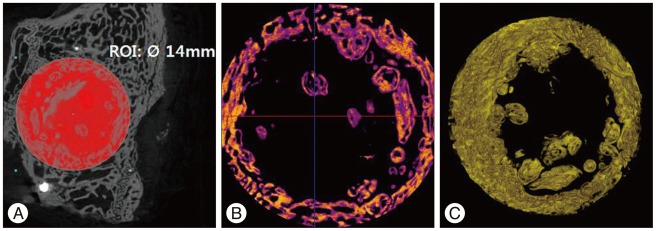
Fig. 5 We defined the region of interest (ROI) as the region within a 14-mm diameter from center point of bone flap. We measured the bone volume (VOLROI) and the bone surface (SURROI) of ROI (A). From VOLROI and SURROI, the bone volume (VOLbone flap) and the bone surface (SURbone flap) of the bone flap, and the volume (VOLmetal) and the surface (SURmetal) of metal fixture were removed. Afterwards, the computed tomography scan images (B) and the 3D reconstruction images (C) of the new bone volume (VOLnew) and the new bone surface (SURnew) were quantified and measured.
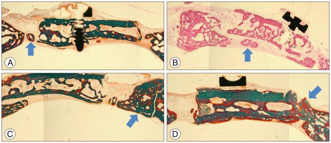
Fig. 6 The histological patterns of new bone formation. The new bone formation was observed in the microscopic examination of the hematoxylin and eosin stain and Goldner's stain in all of the rabbit tissues. The new bone formation was observed as a form of 1) a bony islet (arrow) between the bone flap and the edge of the cranial bone (A), 2) a bony islet (arrow) at the lower part of the bone flap (B), 3) a new bone formation (arrow) at the edge of the cranial bone (C), and 4) a bone flap incorporation (arrow) between the bone flap and the edge of the cranial bone (D).
Reference
-
1. Abbott KH. Use of frozen cranial bone flaps for autogenous and homologous grafts in cranioplasty and spinal interbody fusion. J Neurosurg. 1953; 10:380–388. PMID: 13070062.2. Ahmadi SA, Meier U, Lemcke J. Detailed long-term outcome analysis after decompressive craniectomy for severe traumatic brain injury. Brain Inj. 2010; 24:1539–1549. PMID: 20973624.
Article3. Asano Y, Ryuke Y, Hasuo M, Simosawa S. [Cranioplasty using cryopreserved autogenous bone]. No To Shinkei. 1993; 45:1145–1150. PMID: 8123304.4. Chang V, Hartzfeld P, Langlois M, Mahmood A, Seyfried D. Outcomes of cranial repair after craniectomy. J Neurosurg. 2010; 112:1120–1124. PMID: 19612971.
Article5. Chibbaro S, Di Rocco F, Mirone G, Fricia M, Makiese O, Di Emidio P, et al. Decompressive craniectomy and early cranioplasty for the management of severe head injury : a prospective multicenter study on 147 patients. World Neurosurg. 2011; 75:558–562. PMID: 21600512.
Article6. Chun HJ, Yi HJ. Efficacy and safety of early cranioplasty, at least within 1 month. J Craniofac Surg. 2011; 22:203–207. PMID: 21233757.
Article7. Coutinho JM, Majoie CB, Coert BA, Stam J. Decompressive hemicraniectomy in cerebral sinus thrombosis : consecutive case series and review of the literature. Stroke. 2009; 40:2233–2235. PMID: 19372443.
Article8. De Bonis P, Pompucci A, Mangiola A, D'Alessandris QG, Rigante L, Anile C. Decompressive craniectomy for the treatment of traumatic brain injury : does an age limit exist? J Neurosurg. 2010; 112:1150–1153. PMID: 19645536.
Article9. DeLuca L, Raszewski R, Tresser N, Guyuron B. The fate of preserved autogenous bone graft. Plast Reconstr Surg. 1997; 99:1324–1328. PMID: 9105359.
Article10. Dorfer C, Frick A, Knosp E, Gruber A. Decompressive hemicraniectomy after aneurysmal subarachnoid hemorrhage. World Neurosurg. 2010; 74:465–471. PMID: 21492596.
Article11. Elliott H, Scott HJ. The bone-bank in neurosurgery. Br J Surg. 1951; 39:31–34. PMID: 14858821.
Article12. Elsalanty ME, Genecov DG. Bone grafts in craniofacial surgery. Craniomaxillofac Trauma Reconstr. 2009; 2:125–134. PMID: 22110806.
Article13. Frassanito P, Massimi L, Caldarelli M, Tamburrini G, Di Rocco C. Complications of delayed cranial repair after decompressive craniectomy in children less than 1 year old. Acta Neurochir (Wien). 2012; 154:927–933. PMID: 22198327.
Article14. Gooch MR, Gin GE, Kenning TJ, German JW. Complications of cranioplasty following decompressive craniectomy : analysis of 62 cases. Neurosurg Focus. 2009; 26:E9. PMID: 19485722.15. Grant GA, Jolley M, Ellenbogen RG, Roberts TS, Gruss JR, Loeser JD. Failure of autologous bone-assisted cranioplasty following decompressive craniectomy in children and adolescents. J Neurosurg. 2004; 100(2 Suppl Pediatrics):163–168. PMID: 14758944.
Article16. Güresir E, Raabe A, Setzer M, Vatter H, Gerlach R, Seifert V, et al. Decompressive hemicraniectomy in subarachnoid haemorrhage : the influence of infarction, haemorrhage and brain swelling. J Neurol Neurosurg Psychiatry. 2009; 80:799–801. PMID: 19531687.17. Hofmeijer J, Kappelle LJ, Algra A, Amelink GJ, van Gijn J, van der Worp HB, et al. Surgical decompression for space-occupying cerebral infarction (the Hemicraniectomy After Middle Cerebral Artery infarction with Life-threatening Edema Trial [HAMLET]) : a multicentre, open, randomised trial. Lancet Neurol. 2009; 8:326–333. PMID: 19269254.
Article18. Honeybul S. Complications of decompressive craniectomy for head injury. J Clin Neurosci. 2010; 17:430–435. PMID: 20181482.
Article19. Hutchinson PJ, Corteen E, Czosnyka M, Mendelow AD, Menon DK, Mitchell P, et al. Decompressive craniectomy in traumatic brain injury : the randomized multicenter RESCUEicp study (www.RESCUEicp.com). Acta Neurochir Suppl. 2006; 96:17–20. PMID: 16671415.20. Im SH, Jang DK, Han YM, Kim JT, Chung DS, Park YS. Long-term incidence and predicting factors of cranioplasty infection after decompressive craniectomy. J Korean Neurosurg Soc. 2012; 52:396–403. PMID: 23133731.
Article21. Iwama T, Yamada J, Imai S, Shinoda J, Funakoshi T, Sakai N. The use of frozen autogenous bone flaps in delayed cranioplasty revisited. Neurosurgery. 2003; 52:591–596. discussion 595-596. PMID: 12590683.
Article22. Jaberi J, Gambrell K, Tiwana P, Madden C, Finn R. Long-term clinical outcome analysis of poly-methyl-methacrylate cranioplasty for large skull defects. J Oral Maxillofac Surg. 2013; 71:e81–e88. PMID: 23351772.
Article23. Jüttler E, Schwab S, Schmiedek P, Unterberg A, Hennerici M, Woitzik J, et al. Decompressive Surgery for the Treatment of Malignant Infarction of the Middle Cerebral Artery (DESTINY) : a randomized, controlled trial. Stroke. 2007; 38:2518–2525. PMID: 17690310.
Article24. Klinger DR, Madden C, Beshay J, White J, Gambrell K, Rickert K. Autologous and acrylic cranioplasty : a review of 10 years and 258 cases. World Neurosurg. 2014; 82:e525–e530. PMID: 24036124.25. Matsuno A, Tanaka H, Iwamuro H, Takanashi S, Miyawaki S, Nakashima M, et al. Analyses of the factors influencing bone graft infection after delayed cranioplasty. Acta Neurochir (Wien). 2006; 148:535–540. discussion 540. PMID: 16467959.
Article26. Misch CE. Implantología Contemporánea. Madrid: Mosby;1995. p. 324–350.27. Morina A, Kelmendi F, Morina Q, Dragusha S, Ahmeti F, Morina D, et al. Cranioplasty with subcutaneously preserved autologous bone grafts in abdominal wall-Experience with 75 cases in a post-war country Kosova. Surg Neurol Int. 2011; 2:72. PMID: 21697987.
Article28. Movassaghi K, Ver Halen J, Ganchi P, Amin-Hanjani S, Mesa J, Yaremchuk MJ. Cranioplasty with subcutaneously preserved autologous bone grafts. Plast Reconstr Surg. 2006; 117:202–206. PMID: 16404268.
Article29. Murthy JM, Chowdary GV, Murthy TV, Bhasha PS, Naryanan TJ. Decompressive craniectomy with clot evacuation in large hemispheric hypertensive intracerebral hemorrhage. Neurocrit Care. 2005; 2:258–262. PMID: 16159072.
Article30. Odom GL, Woodhall B, Wrenn FR. The use of refrigerated autogenous bone flaps for cranioplasty. J Neurosurg. 1952; 9:606–610. PMID: 13000480.
Article31. Osawa M, Hara H, Ichinose Y, Koyama T, Kobayashi S, Sugita Y. Cranioplasty with a frozen and autoclaved bone flap. Acta Neurochir (Wien). 1990; 102:38–41. PMID: 2305650.
Article32. Piedra MP, Ragel BT, Dogan A, Coppa ND, Delashaw JB. Timing of cranioplasty after decompressive craniectomy for ischemic or hemorrhagic stroke. J Neurosurg. 2013; 118:109–114. PMID: 23140156.
Article33. Polin RS, Shaffrey ME, Bogaev CA, Tisdale N, Germanson T, Bocchicchio B, et al. Decompressive bifrontal craniectomy in the treatment of severe refractory posttraumatic cerebral edema. Neurosurgery. 1997; 41:84–92. discussion 92-94. PMID: 9218299.
Article34. Prolo DJ, Burres KP, McLaughlin WT, Christensen AH. Autogenous skull cranioplasty : fresh and preserved (frozen), with consideration of the cellular response. Neurosurgery. 1979; 4:18–29. PMID: 450211.35. Schuss P, Vatter H, Marquardt G, Imöhl L, Ulrich CT, Seifert V, et al. Cranioplasty after decompressive craniectomy : the effect of timing on postoperative complications. J Neurotrauma. 2012; 29:1090–1095. PMID: 22201297.
Article36. Sobani ZA, Shamim MS, Zafar SN, Qadeer M, Bilal N, Murtaza SG, et al. Cranioplasty after decompressive craniectomy : an institutional audit and analysis of factors related to complications. Surg Neurol Int. 2011; 2:123. PMID: 22059118.
Article37. Sohn JY, Park JC, Um YJ, Jung UW, Kim CS, Cho KS, et al. Spontaneous healing capacity of rabbit cranial defects of various sizes. J Periodontal Implant Sci. 2010; 40:180–187. PMID: 20827327.
Article38. Stefini R, Esposito G, Zanotti B, Iaccarino C, Fontanella MM, Servadei F. Use of "custom made" porous hydroxyapatite implants for cranioplasty : postoperative analysis of complications in 1549 patients. Surg Neurol Int. 2013; 4:12. PMID: 23493459.
Article39. Stevenson S, Li XQ, Davy DT, Klein L, Goldberg VM. Critical biological determinants of incorporation of non-vascularized cortical bone grafts. Quantification of a complex process and structure. J Bone Joint Surg Am. 1997; 79:1–16. PMID: 9010181.
Article40. Stiver SI. Complications of decompressive craniectomy for traumatic brain injury. Neurosurg Focus. 2009; 26:E7. PMID: 19485720.
Article41. Sultan SM, Davidson EH, Butala P, Schachar JS, Witek L, Szpalski C, et al. Interval cranioplasty : comparison of current standards. Plast Reconstr Surg. 2011; 127:1855–1864. PMID: 21532415.42. Vahedi K, Hofmeijer J, Juettler E, Vicaut E, George B, Algra A, et al. Early decompressive surgery in malignant infarction of the middle cerebral artery : a pooled analysis of three randomised controlled trials. Lancet Neurol. 2007; 6:215–222. PMID: 17303527.
Article
- Full Text Links
-
- Actions
-
Cited
- CITED
-
- Close
- Share
-
- Similar articles
-
- Cranioplasty Using Frozen Autologous Bone
- Analysis of Cranioplasty Using Frozen Autologous Bone Following Post-Traumatic Decompressive Craniectomy
- Delayed and recurrent surgical site infection from resorbed bone fragment after autologous cranioplasty: a case report
- Bone regeneration effects of human allogenous bone substitutes: a preliminary study
- Bone Flap Changes after Cranioplasty Using Frozen Autologous Bone Flaps: A Three-Dimensional Volumetric Reconstruction Study
