J Korean Orthop Assoc.
2007 Dec;42(6):789-794. 10.4055/jkoa.2007.42.6.789.
A Biomechanical Analysis on Disc Pressure Distribution Changes with Interspinous Spinal Spacer Insertion for Lumbar Spinal Stenosis
- Affiliations
-
- 1Korea Health Industry Development Institute, Seoul, Korea.
- 2Department of Biomedical Engineering, Inje University, Gimhae, Korea.
- 3Department of Biomedical Engineering, Konkuk University, Seoul, Korea.
- 4Department of Orthopedic Surgery, Cheil Orthopedic Hospital, Seoul, Korea. shinkcos@nate.com
- KMID: 1947786
- DOI: http://doi.org/10.4055/jkoa.2007.42.6.789
Abstract
-
PURPOSE: To assess the biomechanical effects and effectiveness of an interspinous spinal spacer (ISS) on the intradiscal pressure using in vitro biomechanical tests.
MATERIALS AND METHODS
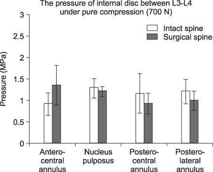
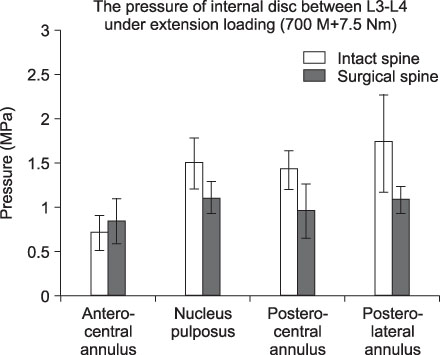
Six calf spine specimens (less than 2 weeks of age, L1-L5) were divided to two groups the intact and the surgery groups (n=3 each). For the surgery group, an ISS made from PMMA (Greek pi=12-mm) were inserted into the space between the spinous processes of L3-L4. The intradiscal pressures at the various regions of the annulus (anterior, posterior, and posterolateral locations) and the nucleus pulposus were measured using the four pressure transducers under pure compression (700 N) and extension loads (700 N+7.5 Nm).
RESULTS
An increase in pressure was observed from neutral to extension at the posterior and posterolateral annulus. After inserting the ISS, the changes in pressure at the adjacent disc levels (L2-L3, L4-L5) were negligible regardless of the loading conditions (p>0.05). However, at the implanted level (L3-L4) statistically significant changes in the pressure were found under extension loading at the nucleus pulposus, posterior and posterolateral regions of the annulus with a pressure drop from 1.48 MPa, 1.42 MPa, 1.71 MPa to 1.11 MPa, 0.961 MPa, 1.08 MPa, at the respective locations (p<0.05). The relative percentage decrease were 25%, 31.7%, and 36.8%.
CONCLUSION
On the implanted level, these results showed that the insertion of the ISS with PMMA can effectively reduce the intradiscal pressures by at least 25% quite uniformly over the intravertebral disc during extension. More effective reduction was observed at the posterolateral location. The pressure changes at the adjacent levels were negligible in contrast to the abnormal pressure changes that are frequently reported after conventional rigid fusion. This suggests that the likelihood of adjacent level degeneration after surgery can be minimized using the ISS insertion.
Figure
Reference
-
1. Adams MA. Mechanical testing of the spine. An appraisal of methodology, result, and conclusion. Spine. 1995. 20:2151–2156.2. Cripton PA, Dumas GA, Nolte LP. A minimally disruptive technique for measuring intervertebral disc pressure in vitro: application to the cervical spine. J Biomech. 2001. 34:545–549.
Article3. Edwards WT, Ordway NR, Zheng Y, McCullen G, Han Z, Yuan HA. Peak stress observed in the posterior lateral anulus. Spine. 2001. 26:1753–1759.4. Gu WY, Mao XG, Rawlins BA, et al. Streaming potential of human lumbar anulus fibrosus is anisotropic and affected by disc degeneration. J Biomech. 1999. 32:1177–1182.
Article5. Kahanovitz N, Arnoczky SP, Levine DB, Otis JP. The effects of internal fixation on the articular cartilage of unfused canine facet joint cartilage. Spine. 1984. 9:268–272.
Article7. Lindsey DP, Swanson KE, Fuchs P, Hsu KY, Zucherman JF, Yerby SA. The effects of an interspinous implant on the kinematics of the instrumented and adjacent levels in the lumbar spine. Spine. 2003. 28:2192–2197.
Article8. Lu YM, Hutton WC, Gharpuray VM. Can variations in intervertebral disc height affect the mechanical function of the disc? Spine. 1996. 21:2208–2216.
Article9. Nagata H, Schendel MJ, Transfeldt EE, Lewis JL. The effects of immobilization of long segments of the spine on the adjacent and distal facet force and lumbosacral motion. Spine. 1993. 18:2471–2479.
Article10. Shono Y, Kaneda K, Abumi K, McAfee PC, Cunningham BW. Stability of posterior spinal instrumentation and its effects on adjacent motion segments in the lumbosacral spine. Spine. 1998. 23:1550–1558.
Article11. Steffen T, Baramki HG, Rubin R, Antoniou J, Aebi M. Lumbar intradiscal pressure measured in the anterior and posterolateral annular regions during asymmetrical loading. Clin Biomech (Bristol, Avon). 1998. 13:495–505.
Article
- Full Text Links
- Actions
-
Cited
- CITED
-
- Close
- Share
- Similar articles
-
- Preliminary Report on Usefulness of Adjacent Interspinous Stabilization using Interspinous Spacer Combined with Posterior Lumbosacral Spinal Fusion in Degenerative Lumbar Disease
- Benefits and Weaknesses of Interspinous Devices in Elderly Patients with Lumbar Spinal Stenosis: Comparative Study of Interspinous U and Decompression Surgery Alone
- Dynamic Stabilization with an Interspinous Process Device (the Wallis System) for Degenerative Disc Disease and Lumbar Spinal Stenosis
- A Morphometric Study of the Lumbar Interspinous Space in 100 Stanford University Medical Center Patients
- Real world adverse events of interspinous spacers using Manufacturer and User Facility Device Experience data


